Download
1 / 31
320 likes | 412 Vues
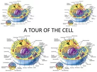
A Tour of the Cell. Chapter 4. The Microscopic World of Cells. Your body is a cooperative society of trillions of cells of many different specialized types. Everything you do reflects processes occurring at the cellular level. Cells that Cure. Heart Attack – lack of oxygen

E N D
A Tour of the Cell Chapter 4
The Microscopic World of Cells • Your body is a cooperative society of trillions of cells of many different specialized types. • Everything you do reflects processes occurring at the cellular level.
Cells that Cure • Heart Attack – lack of oxygen • Do heart muscles regenerate? • Cells – the basic building block of life • Cell Therapy Figure 4.1
The Microscopic World of Cells • Organisms are either: Single-celled - most bacteria and protists Multicelled - plants, animals, and most fungi How do we study this microscopic world?
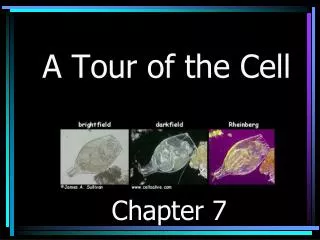
Light Microscope (LM) • Light microscope – visible light passes through a specimen - lenses enlarge, or magnify, the image • 2 important factors: 1) Magnification – increase in the object’s apparent size 2) Resolving power – ability of an optical instrument to show 2 objects as being separate
Cell Theory • Cells were first discovered in 1665 by Robert Hooke - used a microscope to examine a thin slice of cork • By the mid-1800s accumulation of scientific evidence led to the cell theory: - All living thing are composed of cells - All cells are formed from previously existing cells
Electron Microscope (EM) • Uses a beam of electrons - has a higher resolving power than the LM • The EM can magnify up to 100,000X - can distinguish objects as small as 0.2nm - a period at the end of a sentence is 10^6 larger - such power reveals the details of diverse parts in a cell • Biologists began using EMs in the 1950s - a giant leap forward in our understanding of cell structure
Reference measurements along the left side marks a 10-fold decrease in size • Most cells are between 1 and 100um in diameter Fig 4.3
Electron Miscroscopes • Biologists usea SEM to study the detailed architecture of the surface of a cell • The transmission electron microscope (TEM) can be used to explore the internal structure of a cell So, why don’t we just use electron microscopes?
Preparation of a specimen for EM, requires killing and preserving cells before they can be examined Fig 4.2
The 2 Major Categories of Cells • The countless cells that exist on earth fall into basic categories: - Prokayotic cells (bacteria and archea) - Eukaryotic cells (all other organisms – protists, plants, fungi, and animals) • Prokaryotic and eukaryotic cells differ in several important aspects.
Prokaryotic Cells • Are structurally simpler - 1st prokaryotes appeared on Earth over 3.5 bya - 1st eukaryotes did not appear until ~2.1 bya • Are usually much smaller - about 1/10 the length of a typical eukaryotic cell • Lack internal structures surrounded by membranes - eukaryotic cells have several membrane-enclosed organelles • Lack a nucleus – DNA is coiled in a nucleoid region - eukaryotic cells enclose their DNA within a double-membrane
Idealized Prokaryotic Cell Figure 4.5
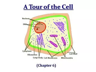
Eukaryotic Cell Similarities • Plasma membrane (PM) – thin outer membrane - regulates traffic of molecules between the cells and their surroundings • Nucleus – membrane enclosed organelle that contains the cell’s DNA • Cytoplasm – region between the nucleus and the PM - consists of organelles suspended in a fluid, the cytosol
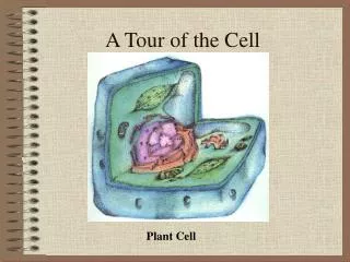
Eukaryotic Cell Differences • Chloroplasts in plant cells - convert light energy to the chemical energy of food • Protective cell wall outside of the PM
Checkpoint • Microscopes – what is the difference between a light microscope and and an electron microscope • Which type would you use to study: a) changes in the shape of a living human white blood cell b) finest details of surface texture of a human hair c) detailed structure of an organelle in the cytoplasm of a human cell • Why would you pick a light microscope?
Checkpoint • How is the nucleoid region of a prokaryotic cell different from the nucleus of an eukaryotic cell • In what type of eukaryotic cell would you find a cell wall. What is its purpose. • What is a chloroplast and where would you find one.
Plasma Membrane: A Fluid Mosaic • PM is the boundary that separates the living cell from its nonliving environment - composed mostly of lipids and proteins • The lipids belong to a special category called phospholipids - related to triglycerides but contain only 2 fatty acid chains - contain a phosphate group • Phospholipids form a two-layered membrane - a phospholipid bilayer
At an interface between 2 aqueous environments: • Phosphate group, electrically charged – hydrophilic • Fatty acid chains are hydrophobic Fig 4.7a
Membrane Proteins • Embedded in the phospholipid bilayer of most membranes are specific proteins that perform various functions • Membrane proteins, like the phospholipids, have both hydrophilic and hydrophobic regions • Fluid Mosaic Model - phospholipid bilayer is not static - Fluid: molecules can move freely within the membrane - Mosaic: a diversity of proteins exists within the membrane
Fluid Mosaic Model of Membrane Membrane Proteins • Have hydrophilic and hydrophobic regions • Drift within the membrane Figure 4.7b
Cell Surfaces • Most cells secrete materials for coats of one kind or another that are external to the PM • These extracellular coats help protect and support cells - and facilitate interactions between cellular neighbors in tissues
Plant Cells • Plant cells have cell walls - help protect the cells - maintains their shape - keep cells from absorbing too much water • Cell walls are made from cellulose fibrils - embedded in a matrix of other molecules • Plant cells are connected to one another via channels that pass through their cell walls - connects the cytoplasm of each cell to its neighbors - allows water and small molecules to move between cells
Animal Cells • Lack a cell wall but most secrete a sticky coat called the extracellular matrix (ECM) - helps hold cells together in tissues - protects and supports the cells • Cells are bound to the ECM by surface proteins in the PM • Surface of most cells contain cell junctions - specialized structure that connect cells to each other - allow adjacent cells to function in a coordinated manner
Checkpoint • How do phospholipids organize themselves in an aqueous solution? Why? • Explain each word in the term fluid mosaic and how this describes the structure of a membrane. • What polysaccharide is the primary component of plant cell walls? • What are the functions of a cell wall? Name its counterpart in an animal cell.