Download

1 / 22
220 likes | 263 Vues
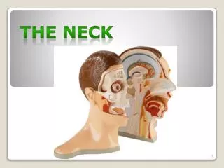
The Neck. Lecture Objectives. Define the boundaries of the neck. Describe the fasciae of the neck. Summarize the main arteries, veins, nerves and lymph nodes of the neck. List the muscles of the neck. Describe the neck triangles.

E N D
LectureObjectives • Define the boundaries of theneck. • Describe the fasciae of theneck. • Summarize the main arteries, veins, nerves and lymph nodes of the neck. • List the muscles of theneck. • Describe the neck triangles. • Describe the key muscles creating the triangles, mainly, sternomastoid, omohyoid anddigastric. • Study the boundaries and content of eachtriangle.
Boundaries of theNeck • Superiormargin • Inferiormargin • Root of theneck
Cervical VertebralMuscles • Suboccipital and deepneck muscles • Mainly extends head andneck • Rectus capitis posterior majorm. • Rectus capitis posterior minorm. • Oblique capitis inferiorm. • Oblique capitis superiorm.
Cervical PrevertebralMuscles • Anterior vertebral muscles • Mainly flexes headand neck • Longus colim. • Longus capitism. • Rectus capitis anteriorm. • Anterior scalenem. • Lateral vertebralmuscles • Mainly laterally flex head andneck • Rectus capitis lateralism. • Splenius capitism. • Levator scapulaem. • Middle scalenem. • Posterior scalenem.
Cervicalfascia • Superficialfascia • Deepfascia: • Investinglayer • Pretracheallayer • Prevertebrallayer • Carotidsheath
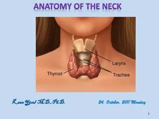
CervicalFascia • Superficial fascia • platysma • Deep fascia: • Investinglayer • Trapezius &SCM • Pretracheallayer • Visceral part ‐ Thyroid & parathyroid • Buccopharyngeal fascia ‐ pharynx • Muscular part ‐ Infrahyoid mm.
CervicalFascia • Deep fascia (continue…) • Prevertebrallayer • Posterior to pharynx & esophagus • Vertebralmm. • Continue as axillarysheath • Carotidsheath • Thickening of the otherlayers • Common & internal carotid aa. • Intrnal jugularv. • Vagusn. • Deep cervical lymphnodes
CervicalFascia: Extensions • Alarfascia • Division from prevertebralfascia • From skull to T2 (merge with buccopharyngeal fascia) • Buccopharyngealfascia • Superior & posterior continuation of the pretrachealfascia
Cervical Fascia:Spaces • Retropharyngealspace • Between buccopharyngeal fascia and prevertebralfascia • Spread ofinfections • (Real) Retropharyngealspace • Between the alar fascia and buccopharyngealfascia • Allow movement of pharynx, larynx, and trachea duringswallowing • Continuous with superior mediastinum to T2 • Danger space • Between the alar fascia and the prevertebralfascia • Continuous withmediastinum
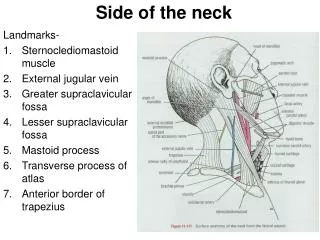
Neck Triangles:Boundaries • Anteriortriangle • Anterior toSCM • Carotidtriangle • Digastrictriangle • Submentaltriangle • Muscular triangle • Posteriortriangle • Posterior toSCM • Anterior totrapezius • Occipitaltriangle • Supraclaviculartriangle
Principal groups of lymph nodes Lymph drainage of Head and neck • Regional lymphnodes • Occipital • Retroauricular • Parotid • Buccal • Submandibular • Submental • Anteriorcervical • Laryngeal • Tracheal • Superficialcervical • Deepcervical • Jugulartrunk
OccipitalTriangle • Above the inferior bellyof omohyoid m. • Spinal accessory nerve(XI) • Junction of the superior & middle thirds of the posterior border of SCM → junction between middle & lower thirds of the anterior border of trapezius • Injury
AnteriorTriangle • Carotidtriangle • Carotidsheath • Between sternocalvicular joint and the mid point between mastoid and angle ofmandible • Hypoglossalnerve • Cervical sympathetictrunk • Submandibular (digastric) triangle • Submandibular gland • Submandibular lymph nodes
Surface Anatomy ofthe Neck • Hyoid bone –C3 • Posterior to themandible • Laryngeal prominence (Adma’s apple)‐ tip(C4) • Vocal cords – at themiddle • Cricoid cartilage –C6 • Cricothyroidligament • Cricothyrotomy • First trachealcartilage • Tracheostomy • Thyroidgland • Isthmus – 2nd – 4th tracheal rings