Download
1 / 41
510 likes | 1.11k Vues
PATENT DUCTUS ARTERIOSUS Neo Case Presentation. By: Socorro Quimson RNC-NIC, BSN, SNNP GNRS 5632 NNP II. OBJECTIVES. Review Maternal/ Infant risk and complications Review Admission Exam, clinical manifestation and diagnostic evaluation Discuss Hospital Course by System and medications

E N D
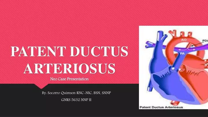
PATENT DUCTUS ARTERIOSUS Neo Case Presentation By: Socorro Quimson RNC-NIC, BSN, SNNP GNRS 5632 NNP II
OBJECTIVES • Review Maternal/ Infant risk and complications • Review Admission Exam, clinical manifestation and diagnostic evaluation • Discuss Hospital Course by System and medications • Describe the primary admission diagnoses, pathophysiology and associated problems • Examine the Pertinent Theories and Evidence based practice • Discuss Family interactions and Discharge follow-up
Maternal History • 28 years old , Married, Hispanic • G 8, P6, Ab 2 • No Prenatal Care • Father of the infant is involve • No record of LMP and EDC • Presented with vaginal bleeding , premature contractions and abdominal pain prior to delivery. She was found to be fully dilated with breech presentation and heavy vaginal bleeding. Taken for stat C-section for placental abruption.
Maternal Labs • Blood Type O positive • RPR Non reactive • HIV Negative • HBsAg Negative • GBS Unknown • Gonorrhea Unknown • Chlamydia Unknown • Rubella Immune
Maternal and Fetal Risks & Complications Maternal • Caesarian Delivery • Placental Abruption • Vaginal Bleeding Infant • Prematurity • Very Low Birth Weight • Respiratory Problems -Pulmonary Edema -Chronic lung Disease • Infections -Septicemia -Necrotizing Enterocolitis • Congestive Heart Failure
Delivery and Stabilization • Infant was delivered via Stat C-section for abruption placenta, and breech presentation • Artificial rupture of membrane at the delivery . With clear amniotic fluid. • Infant was immediately placed on the radiant warmer, dried , suctioned mouth and nose. • Was hypotonic with minimal respiratory effort, Heart rate was below 100 beats/min. • Intubated with 2.5 ETT and PPV was delivered. • Surfactant was administered in the delivery room. • Apgars were 4 and 7 at 1 and 5 minutes of life. • Infant was transported to NICU and placed on SIMV mode of mechanical ventilation with good response.
Admission Assessment and Diagnostics Admission vitals & Measurements: BP: 52/30 ( 32) , T : 98.2 , HR : 168 , Weight : 1340 grams, FOC : 29 cm Birthweight : 960 grams General : Active female infant under radiant warmer. Currently weigh 1340 grams. Skin : Pink , good skin turgor, brisk capillary refill. No evidence of skin lesions. HEENT : Anterior fontanelle is soft and flat, sutures approximated, moderate amount of molding present . Neck is supples, no nodules. With OGT and ETT in place in the mouth at 7.5 cm lip level. Lips and mouth mucosa are pink and intact. Unable to palpate the palate due to the ETT tube in place. Eyes, Nose and Ears are in appropriate size and in normal position. Respiratory : On HFOV, with equal and good chest wiggle. Clear and equal Breath sounds. Heart : Normal rate and rhythm, with III/VI murmur at left upper sternal border. Brachial and femoral pulses palpable.
Admission Assessment and Diagnostics GI : Abdomen is soft, nondistended, no evidence of visceromegaly. Bowel sounds auscultated. Umbilicus is healed. Neuro: Tone appropriate for gestational age MS : Move all extremities. PICC line in Right upper extremity. X-ray showed tip of the catheter positioned at mid-clavicle. Trunk & Spine: Infant’s back appear symmetrical, spine is palpable all along its length. Genitaila: Normal female genitalia, appropriate for gestational age. Anus : Anal opening and wink is present.
Primary Admission Diagnosis • Former 27 week , AGA , now Day of life 30, transferred from other facility for treatment of PDA and ligation evaluation • Primary Diagnosis Persistent PDA • Subsequent Diagnosis Chronic Lung Disease Prematurity Infectious disease evaluation ( History of positive blood culture)
Etiology and Pathophysiology of Primary Admission Diagnosis • Historical information • Galen initially described the ductusarteriosus in the early first century. • Harvey undertook further physiologic study in fetal circulation. • On1888 Munro conducted the dissection and ligation of the ductusarteriosus in an infant cadaver, and it would be another 50 years before Robert E. Gross successfully ligated a patent ductusarteriosus (PDA) in a 7-year-old child. • This was a landmark event in the history of surgery and heralded the true beginning of the field of congenital heart surgery. • Catheter-based closure of the structure was first performed in 1971. • (Kim, 2012, p. 1)
Etiology and Pathophysiology of Primary Admission Diagnosis EPIDEMIOLOGY • Term infant : 0.02% and 0.006% estimated incidence of live births. • Increase incidence to PREMATURE INFANTS and children with a history of perinatal asphyxia, and, possibly, children born at high altitude. Premature infants > 32 weeks' gestation : 20 % < 28 weeks' gestation : 60 % • Low birth weight infants (< 2500 g) : 30 % • Siblings also have an increased incidence. • Perinatal asphyxia usually only delays the closure of the ductus, and, over time, the ductus typically closes without specific therapy. • As an isolated lesion, patent ductusarteriosus (PDA) represents 5-10% of all congenital heart lesions. It occurs in approximately 0.008% of live premature births (Kim, 2012, p. 1)
Etiology and Pathophysiology of Primary Admission Diagnosis FACTORS ASSOCIATED WITH INCREASED INCIDENCE OF PDA • Prematurity • Respiratory Distress Syndrome and Surfactant Treatment • Fluid Administration • Asphyxia • Congenital Syndrome • High Altitude • Congenital Heart Disease • Genetics • Chromosomal Abnormalities • Other ( Low birth weight, Prostaglandins, Low atmospheric oxygen tension and Hypoxia ) (Gomella, 2009, p. 620) (Kim, 2012, p. 1)
Etiology and Pathophysiology of Primary Admission Diagnosis Anatomy and Pathophysiology • The Ductusarteriosus ( DA) is a large vessel that connects the main pulmonary trunk with the descending aorta. • In the utero , DA diverts blood to the placenta for gas exchange. About 90 % of right ventricular outflow is through the DA . • After birth functional closure of the ductus occurs within hours ( but up to 3-4 days) . • Patency of the fetal ductusarteriosus depends on COX-2 derived PGE2 acting on the EP4 receptor. At birth, reduced PGE2 levels , a consequence of increased PGE2 metabolism, allow ductusarteriosus closure (Smyth & FitzGerald, 2012, p.326) (Gomella, 2009, p. 619-620) (Sadowski, 2010, p. 554)
Etiology and Pathophysiology of Primary Admission Diagnosis Anatomy and Pathophysiology cont.. • Complete anatomic closure with fibrosis and permanent sealing of the lumen takes up to 2-3 weeks. • Patent DuctusArteriosus refers to the failure of the closure process and continued patency of this fetal channel. • If the DA remains patent postnatally and pulmonary vascular resistance falls , blood flow through the DA is from the aorta to the pulmonary artery ( Left to Right Shunting). (Gomella, 2009, p. 619-620) (Zahka & Erenberg, 2011, p. 1261)
Etiology and Pathophysiology of Primary Admission Diagnosis Anatomy and Pathohysiology • Improvement in Oxygenation causes the pulmonary vascular resistance to drop rapidly. Although increased oxygen tension is a potent stimulant of smooth muscle contraction, which should decrease patency , premature infants have an immature response to oxygen, thus the PDA remains open. • Lack of ductal smooth muscle ( ex. In premature infants) prolongs patency. • Prostaglandins inhibit closure of ductus. (Sadowski, 2010, p. 554)
Etiology and Pathophysiology of Primary Admission Diagnosis Anatomy and Pathophysiology • As Pulmonary Vascular Resistance (PVR )falls and Systemic Vascular Resistance ( SVR) rises, a left-to –right shunt via the PDA results in blood flow from the aorta into the pulmonary artery, increasing pulmonary blood flow. The increased pulmonary artery pressure and increased left ventricular pressure and volume lead to bilateral CHF. • Because left-to-right flow is dependent on a drop in PVR , infants with pulmonary disease( ex. Respiratory distress syndrome) will show symptoms when lung disease improves. (Sadowski, 2010, p. 554)
Etiology and Pathophysiology of Primary Admission Diagnosis Common Clinical Presentations (Gomella, 2009, p. 621) (Sadowski, 2010, p. 554) Unexplained metabolic acidosis Respiratory Deterioration Other ( tachypnea, crackles, apneic spells) Radiographic findings consists of normal size or mild cardiomegaly , pulmonary edema, and increased vascularity • Heart murmur • Hyperactive Precordium • Bounding peripheral pulses and increased pulse pressure • Hypotension ( low diastolic blood pressure)
Initial Plan of Care • NPO • TPN through PICC line • Total Fluid of 120 ml/kg/day • Surgical Evaluation • Echocardiogram • Cardiology consult • Chest X-ray on admission • Peripheral Arterial Line placement • Continue HFOV support • Chemistry on admission • CBC with diff. , Blood Culture , urine culture and sputum culture on admission • Coagulation Tests, Type and Screen • MRSA screening • Continue Vancomycin and Cefepime • Continue Versed PRN for agitation • Social Work Consult
Hospital Course by System GESTATION • Former 27 weeks, AGA, now 31 2/7 weeks post conceptional age HEENT • Unremarkable CNS • Head ultrasound on 04/16/2014 , 04/21/2014, 04/29/2014 revealed no obvious bleed in the germinal matrix. Head ultrasound on 05/12/2014 manifested no acute intracranial process , incidental of cavum septum pellucidum( normal anatomic variant). • Head ultrasound follow-up on 05/16/2014.
Hospital Course by System RESPIRATORY SYSTEM • At birth , infant was intubated in the delivery room. Initial diagnosis at birth was RDS and Prematurity. Required Surfactant administration. Was on Mechanical Ventilator from birth to May 9,2014. Failed extubation attempt on 04/15/2014. Was placed on HFOV support for increased episodes of desaturation and hyercapnia on 05/13/2014. Current HFOV setting: Delta P 16, MAP 15, HZ 10, Fi02 55 to 73%. • 05/14/2014 : Chest x-ray shows ETT at T3 about 2 cm above the carina, PICC line tip at mid clavicular line, Lung fields manifests diffuse haziness which can signify pulmonary edema due to left to right shunting via the PDA. Lungs are well expanded at T9. Heart size is unremarkable.
Hospital Course by System CARDIOVASCULAR • Echocardiogram done on 04/16/2014 showed moderate PDA with moderate left to right shunting, qualitative normal cardiac chamber size and function. • Infant received Indocin every 24 hours x3 on 04/18/2014. • Echocardiogram obtained on 04/21/2014 for post Indocin evaluation manifested small PDA. Repeat evaluation done through Echocardiogram on 05/11/2014 showed small to moderate PDA. On admission 05/12/2014 infant’s echocardiogram showed Left Atrial Enlargement and Persistent PDA. • Repeat echocardiogram and Cardiac consultation on 05/13/2014. • PDA LIGATION DONE ON 05/14/2014
Hospital Course by System FLUIDS, ELECTROLYTES, AND NUTRITION • Infant has been on TPN since 04/16/2014 to the present. • 0426/2014 : Started Enteral feeding with Similac Special Care 20 calories every 3 hours. Feeding was advanced up to 15 ml via NGT. • On admission infant placed on D10 TPN via PICC Line at 120 ml/kg/day • 05/13 – 05/14/2014 NPO for possible PDA ligation . • 05/13/2014: Unremarkable Chemistry and liver function test • 05/14/2014 Pre and Post PDA ligation plan : D10 TPN at 120 ml/kg/day
Hospital Course by System HEMATOLOGY • Infant has required multiple PRBC transfusion. Hematocrit on admission 05/13/2014 was 33 mg/dl. • 05/13/2014 : PRBC transfusion 10 ml/kg IV x2. Coagulation test , type and screen prior to transfusion. • Repeat CBC differential on 04/14/2014 . SKIN • 05/14/2014 : Post PDA ligation site at left postero-lateral thorax is clean and dry .
Hospital Course by System MUSCULOSKELETAL • Infant was on breech presentation at birth. There is no evidence of hip dysplasia. INFECTIOUS DISEASE • Maternal GBS status is unknown. Infant received Ampicillin and Gentamicin on 0414/2014 to 04/20/2014. • On 05/02/2014 Blood culture was positive for Staphylococcus Epidermidis. Vancomycin and Cefepime started on 05/02/2014 . • 05/13/2014 : Culture from blood, urine and sputum. Twenty four hour result is negative. • 05/13/2014 : Last dose of antibiotics was given on an hour prior to PDA ligation.
Hospital Course by System GENITOURINARY • Infant has been voiding and stooling. Urine Output within normal limits. HYPERBILIRUBINEMIA • Total Bili peaked at 5.4 on 04/16/2014. Required phototherapy on 04/16/2014 to 04/19/2014. • 05/12/2014 : Total bilirubin level was 0.6 , Direct Bilirubin level was 0.2 ( taken at the transferring facility) • 05/14/2014 : Total and Direct bilirubin. Result still pending. Endocrinology 05/08/2014 : Abnormal newborn screen ( Low T4, T3 and free T4 ) . Thyroid Profile ( Normal)
Medications Infant received the following Medications: • Ampicillin from 04/14/2014 to 04/20/2014 • Gentamicin from 04/14/2014 to 04/20/2014. Levels unremarkable. • Vancomycin from 05/02/2014 to 05/14/2014 • Cefepime from 05/02/2014 to 05/14/2014 • Midazolam prn for agitation • Fentanyl prn for pain • Lasix post PRBC transfusion • Indocin for PDA x 3 doses • Caffeine from 04/15/2014 to 05/05/2014 ( stopped due to tachycardia) • Aldactone and Diuril from 05/10/2014 to 05/12/2014
Pertinent Theories and Evidence Based Practice CONSERVATIVE MANAGEMENT Positive End Expiratory Pressure Diuretics Fluid Restrictions Increasing Hematocrit (Gomella, 2009, p. 621) (Sadowski, 2010, p. 555)
Pertinent Theories and Evidence Based Practice MEDICAL MANAGEMENT INDOMETHACIN • Is a prostaglandin synthetase inhibitor • Has been proven to be effective in promoting ductal closure • It’s effectiveness is limited to premature infants and decreases with increasing postnatal age • Has limited efficacy beyond 3 to 4 weeks of age , even in premature infants. (Gomella, 2009, p. 620)
Pertinent Theories and Evidence Based Practice MEDICAL MANAGEMENT THREE APPROACHES IN ADMINISTERING INDOMETHACIN Prophylactic indomethacin : Indomethacin dose is 0.1 mg/kg/dose IV over 20 minutes every 24 hours from first day of life for 6 days. Early Symptomatic Indomethacin: Infants are given Indomethacin 0.2 mg/kg IV over 20 minutes. Second and third dose are given 12 and 36 hours after first dose. The second and third dose are 0.1 mg/kg/dose if the infant is less than 1250 grams birthweight and less than 7 days old. If the infant is either more than 7 days old or more than 1250 grams the second and third dose are also 0.2 mg/kg /dose. Indomethacin is given if there is any clinical sign of a PDA ( ex. Murmur) and before there are signs of overt failure. Late Symptomatic Indomethacin : Given when signs of congestive failure appear ( usually at 7 to 10 days ). Dosase is 0.25 mg/kg at 12 to 24 hour interval. (Gomella, 2009, p. 620)
Pertinent Theories and Evidence Based Practice COMPLICATIONS OF INDOMETHACIN CONTRAINDICATIONS OF INDOMETHACIN Serum creatinine more than 1.7 mg/dl Frank renal or gastrointestinal bleeding or generalized coagulopathy Necrotizing enterocolitis ( NEC) Sepsis : All inflammatory drugs should be withheld if there is sepsis. Indomethacin may be given once this is under control. Renal Effects • Transient decrease in the glomerular filtration rate and urine output Gastrointestinal Bleeding • Heme-positive stool ( transient) • Indomethacin is a mesenteric vasoconstrictor Platelet Function • Indomethacin impairs platelet function for 7 to 9 days regardless of platelet number. (Clyman, 2012, p. 758) (Gomella, 2009, p. 622
Pertinent Theories and Evidence Based Practice MEDICAL MANAGEMET CONT. IBUPROFEN (NeoProfen) • Another nonselective cyclooxygenase inhibitor, has been shown to close the ductus in animals and preterm infants. • It appears to be as effective as Indomethacin in producing PDA closure in Very Low Birth Weight infant • Does not appear to affect mesenteric blood flow and has less effect on renal perfusion. • Animal studies suggest that ibuprofen may have some cytoprotective effects in the intestinal tract. • Treatment consist of an initial dose ( 10mg/kg infused over 15 minutes) followed by two or more doses ( 5mg/kg each) given 24 and 48 hours later. (Clyman, 2012, p. 759) (Lehne, 2013, p. 898)
Pertinent Theories and Evidence Based Practice SURGICAL TREATMENT • Indicated if a clear contraindication to indomethacin administration exists , or if the drug is ineffective . Relapse after a second short course of indomethacin or after a single day course is also an for surgery. • Standard approach is surgical ligation via posterolateral thoracotomy incision risk. • Surgical mortality rate is less than 1 % (Kennedy, Hoover, Williams, & Iskersky, 2011, p. 691) (Sadowski, 2010, p. 556) (Yates, 2012, p. 644)
Pertinent Theories and Evidence Based Practice PDA IN PRE TERM INFANT • The DA in the preterm infant is thin walled and less dependent on the vasa vasorum, so it does not develop the ischemia-hypoxia-stimulated remodeling unless there is a cessation of blood flow. The preterm ductus is less likely to constrict with birth due to the presence of immature myosin isoforms, potassium channels that are less responsive to oxygen inhibition, altered Rho/Rho kinase pathways, higher circulating PGE2 levels secondary to decreased clearance by the immature lungs, and especially increased sensitivity to the vasodilating effects of PGE2 and Nitric Oxide. (Blackburn, 2013, p. 287)
Pertinent Theories and Evidence Based Practice PDA IN PRE TERM INFANT • The preterm infant has less pulmonary arterial muscle and an immature pulmonary parenchyma. The presence of a patent DA is associated with interstitial edema and decreased compliance of the lung due to pulmonary edema. Ductal closure results in an increased compliance and a decreased need for ventilator y support. • A large left –to-right shunt through the PDA increases the left atrial and ventricular volume, leading to enlargement of these two chambers. The increased left ventricle size also increases the myocardial wall stress, which could lead to myocardial ischemia in the preterm infant. (Blackburn, 2013, p. 287-288)
Pertinent Theories and Evidence Based Practice PDA IN THE FULL TERM INFANT • PDA accounts for 10% of all congenital heart disease in full-term infants . • The PDA in a full term infants is structurally different , which may explain why it does not respond appropriately to the various stimuli for closure. • Indomethacin is usually not effective. The infant should be monitored carefully , and surgical ligation should be considered at the earliest signs of significant congestion. • Even without signs of failure, the PDA should be ligated before one year of age to prevent endocarditis and pulmonary hypertension. (Gomella, 2009, p. 623)
Family Interactions • Parents home is about 180 miles from the facility. • Father was able to travel with transport team. • Father have shown support , care and concern about the infant. • Father was in the NICU waiting area while surgery was ongoing and stayed until surgery was completed. • Mother is currently sick and will not be able to visit.
Discharge Plan and Follow-up • Developmental follow-up • ECI • Cardiac follow-up • Eye exam follow-up • Hearing screening test • Newborn Screening Test • Pediatrician follow-up • Car Seat Challenge Test • Vaccination • CPR class to infant’s primary care giver ( parents) • Social Service Consult to assist the need of the family • Discharge teaching to parents
SUMMARY • The frequency that a premature neonate will develop a hemodynamically significant left-to right shunt through a PDA is inversely proportional to advancing gestational age. • The typical presentations of PDA are harsh systolic ejection murmur heard over the entire precordium, but loudest at the upper sternal border and left infraclavicular areas, as the PVR drops the murmur becomes continuous, bounding peripheral pulses, Wide pulse pressure from 15 to 25 mm hg, patients respiratory status deteriorates, serialchest x-rays show increase in heart size and the lungs may appear radiopaque. • Initial management are fluid restrictions, ventilatory support, and diurIetic therapy. • In symptomatic patients Indomethacin and Ibuprofen are initially used for nonsurgical closure of PDA. • Surgical ligation is indicated for symptomatic patients who do not respond to a second treatment with indomethacin treatment or if indomethacin or ibuprofen treatment is contraindicated
References Blackburn, S. T. (2013). Maternal, fetal, and neonatal physiology : A clinical perspective (4th ed.). Maryland Heights, MO: Elsevier Saunders . Clyman, R. L. (2012). Patent ductusarteriosus in the preterm infant . In C. A. Gleason, & S. U. Devaskar (Eds.), Avery’s diseases of the newborn (9th ed., pp. 751-761). Philadelphia, PA 19103-2899: Elsevier Saunders . Gomella, T. L. (Ed.). (2009). Neonatology: Management, procedures, on-call problems, diseases, and drugs (6th ed.). New York: The McGraw-Hill Companies . Kennedy, P. M., Hoover, D., Williams, L. C., & Iskersky, V. (2011). Cardiovascular diseases and surgical interventions. In S. L. Gardner, B. S. Carter, M. Enzhan-Hines, & J. A. Hernandez (Eds.), Merenstein and Gardner’s handbook of neonatal intensive care (7th edition ed., pp. 678-716). St. Louis, Missour 63043: Mosby Elsevier . Kim, L. K. (2012). Patent ductusarteriosus . Retrieved May 26, 2014, from http://emedicine.medscape.com/article/891096-overview#a0104
References Lehne, R. A. (2013). Pharmacology for nursing care (8th ed.). St. Louis, Missouri 63146: Elsevier Saunders. Sadowski, S. L. (2010). Cardiovascular disorders . In T. Verklan, & M. Walden (Eds.), Core curriculum for neonatal intensive care nursing (4th ed., pp. 534-588). St. Louis, Missouri 63146: Saunders Elsevier . Smyth, E. M., & FitzGerald, G. A. (2012). The eicosanoids: Prostaglandins,thromboxanes, leukotrienes, and related compounds. In B. G. Katzung, S. B. Masters, & A. J. Trevor (Eds.), Basic and clinical pharmacology (12 th ed., pp. 313-329). New York : Mc Graw Hill Medical . Yates, R. W. (2012). Cardiovascular disease . In J. M. Rennie (Ed.), Rennie and Robertson’s textbook of neonatology (5th ed., pp. 617-669). UK : Churchill Livigstone Elsevier . Zahka, K. G., & Erenberg, F. (2011). Congenital defects. In R. J. Martin, A. A. Fanaroff, & M. C. Walsh (Eds.), Fanaroff & Martiin’s neonatal- perinatal medicine diseases of the fetus and infant (9th ed., pp. 1245-1266). St. Louis, Missouri 63043: .