Download
1 / 62
651 likes | 1.19k Vues
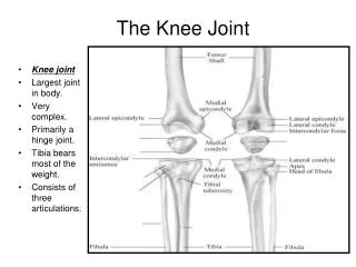
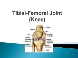
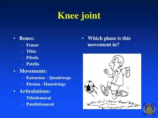
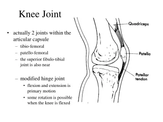
Knee Joint Kinesiology. Amir H. Bakhtiary PhD, PT Associate Professor. Physiotherapy Department Rehabilitation faculty Semnan University of Medical Sciences. Knee Extensors Muscles. Gluteus Max and Soleus may help knee Ext in Closed Kinematic Chain (Movements. Quadriceps.

E N D
Knee Joint Kinesiology Amir H. Bakhtiary PhD, PT Associate Professor Physiotherapy Department Rehabilitation faculty Semnan University of Medical Sciences
Gluteus Max and Soleus may help knee Ext in Closed Kinematic Chain (Movements
Quadriceps عضله چهارسر رانی • تنها بخش دومفصلی آن رکتوس فموریس است • جهت نیروی کشش آن نسبت به تنه فمور • 7-10 درجه به داخل و 3 تا 5 درجه به جلو • جهت کشش VL 35 درجه بطرف خارج • جهت کشش VI موازی با تنه فمور • جهت کشش VM • الیاف فوقانی 15 – 18درجه بطرف داخل • الیاف تحتانی یا مایل 50-55 درجه بطرف داخل • بخش VM درانتهای دامنه Ext زانو فعال می شود
نقش پاتلا بر عملکرد عضله چهار سررانی را تعریف کنید؟ قرار گرفتن پاتلا در داخل تاندون عضله چهار سررانی • افزایش گشتاور نیروی عضله چهارسررانی از طریق • افزایش MA تاندون چهار سررانی • افزایش فاصله تاندون کوادریسپس و پاتلا از محور حرکت زانو • تغییر جهت خط کشش عضله کوادریسپس • افزایش زاویه کشش تاندون • کاهش اصطکاک بین تاندون و سطح فمور • نقشی بیشتر از یک قرقره ساده بازی کرده • هم تغییر جهت نیرو • هم تغییر اندازه نیرو • برداشتن پاتلا موجب کاهش نیروی کوادریسپس تا 49% بدلیل کاهش MA
Increase the muscle force by Knee Ext in OKC Cause More tension on the ACL
Decease the muscle force by Knee Ext in OKC Cause Less tension on the ACL
Patellofemoral Joint نقش پاتلا در مفصل پاتلوفمورال مانند قرقره اکسنتریک آناتومی شامل 1) تغییر جهت نیرو، 2) افزایش اثر نیرو و 3) کاهش اصطکاک بین سطوح • بستگی به قابلیت تحرک چهار گانه آن دارد • Patellar Flexion/Extension (تحرک اصلی) • Patellar Tilt (اجازه تطبیق پاتلا با کندیلهای نامتقارن فمور) • Med Tilt (بین 0-30 الی 100 درجه Flex) • Lat Tilt (بین 20 الی 100 درجه Flex) • Med & Lat Rotation (چرخش زاویه تحتانی پاتلا به تبعیت از تیبیا) • بین 25 الی 100 درجه Flex پاتلا 6-7 درجه به خارج می چرخد • Medio-lateral Translation یا Patellar Shift • Active: در انتهای EXT 7/5 تا 10 mm جابجایی خارجی • Passive: • Full Ext (جابجایی داخلی: 9.6 mm، جابجایی خارجی: 5.4 mm) • در 35درجه Flex (جابجایی داخلی: 9.4 mm، جابجایی خارجی: 10 mm)
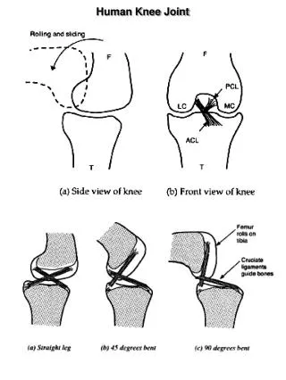
Open kinematics Chain Patella Movement During Knee Flexion
Patella Movement During Knee Flexion Closed kinematics Chain
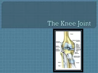
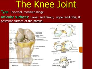
Patellofemoral Joint Surface سطح مفصلی پاتلوفمورال • ناسازگارترین سطوح مفصلی بدن • سطح مفصلی پاتلا بسیار کوچکتر از فمور • خصوصیات غضروفی ان از یک نقطه به نقطه دیگر متفاوت است • سطح مفصلی پاتلا توسط غضروف ضخیمی پوشیده شده • توسط یک ستیغ مرکزی به دو بخش داخلی و خارجی تقسیم شده • هر دو بخش صاف تا کمی محدب (در دو صفحه ساژیتال و فرونتال) • در 30% موارد فاست داخلی تری به نام Odd Facet • سطح مفصلی فمور با یک شیار به دو بخش داخلی و خارجی • در فرونتال مقعر و در ساژیتال محدب (فاست خارجی محدب تر) • زاویه بین دو فاست °138 (از 116 تا ° 151 بین افراد مختلف متغییر)
Patellofemoral Contact Surface سطح تماس پاتلا با فمور • هیچ تماسی در Full ext وجود ندارد • با افزایش Flex تماس سطوح افزایش یافته (از پایین به بالا) • در 10 تا 20 درجه Flex اولین تماس با سطح تحتانی پاتلا • در 45 درجه flex نیمه میانی پاتلا وارد تماس شده • در 90 درجه flex تمام قسمتهای پاتلا وارد تماس شده • با flex بیش از 90 درجه پاتلا وارد شیار بین کندیلی شده و Odd facet در تماس قرار گرفته • در 135 درجه flex تماس فقط در فاست خارجی و Odd Facet • فاست داخلی بیشترین تماس و odd Facet کمترین تماس را تجربه • عدم تعادل در نیروهای وارده بر غضروف مفصلی منجر به تغییرات دژنراتیو در این فاستها می گردد
Reaction Force on the Patellofemoral Joint نیروهای وارده روی مفصل پاتلو فمورال • در Full ext نیروهای کوادریسپس و پاتلا یکدیگر را خنثی کرده • پاتلا در حالت تعادل در مقابل فمور قرار دارد • با افزایش Flex برآیند این دو نیرو موجب فشرده شدن پاتلا روی فمور • با افزایش Flex این نیرو افزایش یافته • موجب نیروی عکس العمل روی سطوح مفصلی گردیده که میزان آن تحت تاثیر • Knee Flexion • Quadriceps Force • Elastic Passive Force • اندازه نیروی عکس العمل • راه رفتن عادی (10 تا 15 درجه Flex زانو) 50% وزن بدن • بالا و پائین رفتن از پله (60 تا 90 درجه Flex زانو) 3.5 برابر وزن بدن • فعالیتهای همراه با Flex شدید زانو (135 درجه Flex) تا 8 برابر وزن بدن
What is the Compensatory Mechanisms for Compressive Force Distribution in patelofemoral joint? • Contact area with knee flexion • Medial facet contact from 30-70 • Thickest hyaline cartilage in body • Largest QF MA 30-70 • QF torque as MA decreases • QF tendon contacts condyles 70-90
What are the Medial and Lateral Stability factors of Patellofemoral Joint? ثبات طرفی مفصل PF تحت تاثیر متقابل دو مکانیسم قرار دارد • مکانیسم عرضی (رتیناکولوم اکستانسوری) • مکانیسم طولی (تاندونهای پاتلا و کوادریسپس) • تعادل بین این دو مکانیسم منجر به حرکت صحیح پاتلا هنگام حرکات زانو میشود یا Patellar Tracking • در صورت بر هم خوردن تعادل بین عوامل فوق حرکت پاتلا روی فمور دچار اختلال می گردد
Normal Patella Tracking • Maintains maximum congruence • Passive restraints • Active restraints
Abnormal Patella Tracking • ↓ congruence • Stretches capsule & retinacula • ↓ contact area Lateral Medial
Causes of Abnormal Tracking • Skeletal abnormalities • Strength imbalance in QF • Strength imbalance in fibrous tissues • Compensatory movements in knee due to abnormal foot movement
Causes of Abnormal Tracking • Skeletal abnormalities • Strength imbalance in QF • Strength imbalance in fibrous tissues • Compensatory movements in knee due to abnormal foot movement
Skeletal Abnormalities: Q-angle مقدار طبیعی = 10 تا °15 با Flex کاهش یافته عوامل موثر بر افزایش آن پهنتر بودن لگن افزایش anteversion فمور افزایش Valgus زانو
Increases in Q-angle are associated with: • femoral anteversion • external tibial torsion • laterally displaced tibial tubercle • genu valgus • Wide Hips (female runners) • Pronation of the feet • Subluxating Patella • High riding patella (Patella Alta) • Weak Vastus Medialis
Skeletal Abnormalities: Genu Varum & Genu Valgum • Q angle w/ age • Varum common in very young children • Valgum seen in growing children • Menisectomy effects
Skeletal Abnormalities: Patella Alta & Patella Baja • Index of Insall & Salviti • LP/LT • Normal = 1.0 • Patella alta = 0.8 • Patella baja = 1.2 • Women ratio
Skeletal Abnormalities: Patella Surface Lateral Border • Appositional forces ↓ in full extension • Prominence of lateral border prevents lateral displacement • Underdevelopment common in children as growing
Lateral tracking Skeletal Abnormalities: Femoral & Tibial Torsion
Causes of Abnormal Tracking • Skeletal abnormalities • Strength imbalance in QF • Strength imbalance in fibrous tissues • Compensatory movements in knee due to abnormal foot movement
Causes of Abnormal Tracking • Skeletal abnormalities • Strength imbalance in QF • Strength imbalance in fibrous tissues • Compensatory movements in knee due to abnormal foot movement
Causes of Abnormal Tracking • Skeletal abnormalities • Strength imbalance in QF • Strength imbalance in fibrous tissues • Compensatory movements in knee due to abnormal foot movement
Compensatory Movement • Pronation of foot accompanied by medial rotation of tibia medial rotation & medial translation of patella • Pronation coupled w/ forceful quadriceps femoris leads to anterior tilt • EX: jumping, landing, running
Meniscal lesion can also occur through a collision and is thought deep knee bends can also be a cause. A transverse tear of the lateral meniscus A transverse tear of the lateral meniscus A longitudinal tear of the lateral cartilage meniscus
Damage to the medial meniscus at it's attachment to the ligament. Also shown a longitudinal tear in the lateral meniscus.The cartilage gets squeezed between the bones with most of your bodyweight on top!