Download
1 / 4
40 likes | 134 Vues
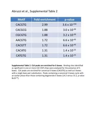
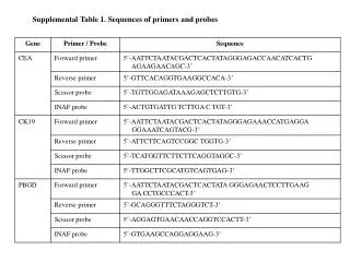
Online Supplemental Table. Online Supplemental Figures. I. I. I. M. M. sirius red. H.E. I. I. M. M. HHF35 (SMC). CD68 (M ). II. I. I. M. M. PEDF. non-immune IgG. I. PEDF. M. Legend for online supplemental figures.

E N D
Online Supplemental Figures I I I M M sirius red H.E. I I M M HHF35 (SMC) CD68 (M) II. I I M M PEDF non-immune IgG I PEDF M
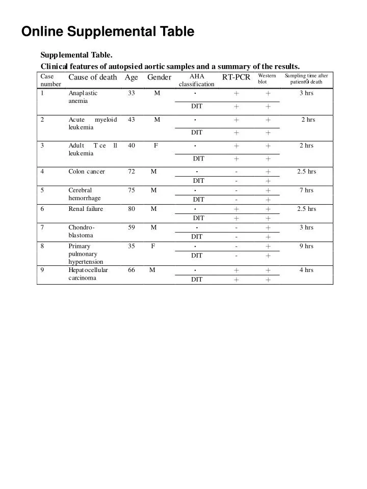
Legend for online supplemental figures. I and II. Histopathological and immunohistochemical findings of grossly non-atherosclerotic human aorta (histological DIT). I: intima and M: media. I. Histology of case 12 (online supplemental table, 31 year-old female). Diffuse collagen deposition was seen in both the intima and the media (upper right). CD68-positive monocytes/macrophages (brown, lower right) were scattered in the diffusely thickened intima, and HHF35-positive smooth muscle cells were ubiquitously located in both the intima and the media (brown, lower right). Original magnification: x20. II. IHC for PEDF in the serial sections of b. The panel at the lower left shows a high-powered view of the boxed area shown in the upper left panel. A diffusely positive reaction was observed in the intima (brown reaction, upper and lower left), and this reaction was scattered in the tunica media (upper left). No positive signal was observed in the tissue sections that had been reacted with isotype-matched non-immune IgG1 (upper right). Original magnification: upper panels, x20; lower left panel, x40.