Download
1 / 74
830 likes | 1.61k Vues
Myelodysplastic, Myeloproliferative, and Histiocytic Disorders. Kenneth McClain M.D. Ph.D. Texas Children’s Cancer Center Houston, TX. Disclosure Information. Own common stock of Johnson & Johnson Co. No discussion of unlabeled uses *=New material not in syllabus.

E N D
Myelodysplastic, Myeloproliferative, and Histiocytic Disorders Kenneth McClain M.D. Ph.D. Texas Children’s Cancer Center Houston, TX
Disclosure Information • Own common stock of Johnson & Johnson Co. • No discussion of unlabeled uses*=New material not in syllabus
What is Myelodysplastic Syndrome (MDS)or… When Do Blasts in the Marrow Not = Leukemia? Pediatric version of WHO Criteria for MDS • Absence of AML cytogenetic findings • Two or more of the following:Sustained cytopeniaDysplasia in 2 cell linesClonal cytognenetic abnormality (5q-, monosomy 7)5-19% Blasts (>20% Blasts = AML)
MDS Can Become AML,But is not AML a priori • May need several marrow exams to establish diagnosis of MDS vs. AML • Incidence of MDS ~ 1.5 per million10-20% become AML
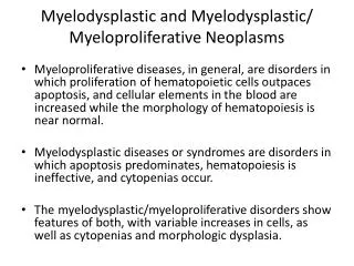
Pediatric MDS Classification Three major categories:1. Adult-Type Myelodysplastic Syndromes2. Down Syndrome with abnormal megakaryocyte proliferation3. Myelodysplastic/Myeloproliferative Syndrome: JMML
For Perspective-Adult MDS • Predominant feature: Marrow Failure • Most frequent in adults 40-60 yrs. • Two major clinical groups1. High incidence of progression to AML: Multilineage/Mutator Phenotype2. Low Progression to AML: Unilineage
Types of Adult MDS • High Incidence of progression to AML:Refractory Cytopenia with multilineage dysplasia: (RCMD)Refractory Anemia with excess Blasts (RAEB) • Low Incidence of progression to AML:Refractory AnemiaRefractory anemia with ringed sideroblastsdel 5q: Macrocytic anemia
Pediatric MDS • Often with an underlying condition:Aplastic anemia, Fanconi anemia, platelet storage pool defect, neurofibromatosis, secondary to malignancy treatment Syndromes: Down, Kostmann’s, Shwachman-Diamond, Dyskeratosis congenita, Bloom’s, Noonan’sAmegakaryocytic thrombocytopeniaFamilial monosomy 7, 5q-
Differential Diagnoses of MDS:Need >1 Marrow Finding and Cytogenetic Data • Other anemias:megaloblastic congenital dyserythropoietic sideroblastic anemia • Leukemia/pre-leukemia:Megakaryocytic leuk. Myelofibrosis PNH • Toxins: Arsenic, chemotherapy • Virus: HIV
Myelodysplastic Syndrome (MDS) • Refractory cytopenia (RC): <2% PB blasts,<5% marrow blasts • Refractory anemia with excess blasts (RAEB):2-19% PB blasts, 5-19% marrow blasts • *RAEB in transformation (RAEB-T) PB or marrow blasts 20-29%: Now = AML(Change from Handout) • Marrow abnormalities: 2-3 lineages dysmorphic, erythroid most abnormal
Molecular Genetics of MDS • AML1/RUNX1 gene: point mutationsRegulates hematopoiesis & most frequent translocation in MDSAML • Chromosome 7 & 20 abnormalities in Shwachman synd: “mutator phenotype”
Treatment of MDS • Refractory cytopenia: “expectant follow-up” • RAEB/RAEB-T: ChemotherapyBMT Event-free survival: 14-55% 65-80% (If successful induction)
Down Syndrome Proliferative Diseases • Transient abnormal myelopoiesis (TAM) • Myelodysplastic syndrome (MDS)/acute myeloid leukemia (AML)
DOWN SYNDROMETransient Myeloproliferative Disorder orTransient Abnormal Myelopoiesis • TMD/TAM: leukemoid reaction: usually megakaryocytic • Progression to megakaryocytic leukemia:20%Blasts same in both by morphology, immuno-phenotype GATA-1 *exon 2 mutations in leukemia onlyUltimately clonal cytogenetic data differentiates
Transient Abnormal Myelopoiesis in Down Syndrome MedianRange Age at onset (days) 2 0-180 Hepatosplenomegaly 69% Bruising/petech/bleeding 25% Resp. distress 21% WBC (per l) 47,000 5,000-384,000 Absolute blast ct. 13,000 0-280,000 Hgb (g/dl) 16.8 4-23.2 Platelets (per l) 102,000 5,000-1,800,000
TAM Marrow Characteristics • Hypo- to hypercellular • Fibrosis common • Blasts 32% (range 6.8-80%) • *Immunophenotype: CD7,33,45,34+Platelet markers CD41/42b/61: variably +Best is EM with immunogold labeling of CD61
TAMClinical Outcomes • Onset: median 16 mo. (range 1-30 mo.)No clinical differences between those with or without ANLL • Duration: *Clear blasts median 2 mo., max 6 mo. • *Leukemia 20% (9-38 mo.) 90% M7, rare ALL • 17% died in first few mo. (not leukemia): sepsis, congestive heart failure, hyperviscosity, “crib death”, DIC • But….33% additional hematologic problems: 84% of these developed ANLL Others: CML, MDS, chronic thrombocytopenia
Pediatric MDS Classification:Myelodysplastic/myeloproliferative • Juvenile myelomonocytic leukemia1% of pediatric leukemia cases • Chronic myelomonocytic leukemiaVery uncommon in children • BCR/ABL-negative chronic myelogenous leukemia
Juvenile Myelomonocytic Leukemia JMML • Clinical criteria: hepatosplenomegaly, lymphadenopathy, pallor, fever, skin rash • Minimal lab criteria (need all 3) No t9;22 or bcr/abl rearrangement Peripheral blood monocytosis: >1X10⁹/L Bone marrow blasts <20% (differs from handout)
JMMLAdditional Lab Criteria Need at least 2 of these:-Hgb F increased for age-Myeloid precursors in periph. blood smear-WBC >109/L-Clonal abnormality not always present (monosomy 7, t(5;8), trisomy 8, monosomy 22)-GM-CSF hypersensitivity of monocyte progenitors in vitro-Autonomous growth of CD34+ cells
Molecular Pathogenesis of JMML • Frequent deletions of NF1Negative regulator of Ras signaling • Missense mutations in PTPN11: all Noonan synd. Pts with JMML and 35% of other JMML • Mutations of KRAS2 & NRAS Bottom line: Ras activation central to JMML and other leukemias
Treatment of JMML • Chemotherapy: 16% survival rate @ 3 yrs.Median time diagnosis to death is 15 mo. • Stem cell transplant: 50% survival • *Current COG trial: pre-transplant chemotherapycis-Retinoic acid: inhib “spontanteous outgrowth CFU-GMfludarabine: potentiate metabolism of Ara-C to Ara-CTPAra-C: potent anti-myeloid malignancy therapyfarnesyl protein transferase inhb: anti-Ras*= New data not in syllabus
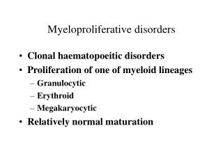
What is a myeloproliferative disorder? • Elevated numbers of a particular cell line in peripheral blood • Hyperplasia of that lineage in the marrow • No secondary causes: infection, drugs, toxins, autoimmune, non-hematologic malignancy, trauma
Types of Myeloproliferative Syndromes • Erythroid: polycythemia vera • Granulocytic: CML • Monocytic: JMML • Megakaryocytic: Essential or familial thrombocytosis, myeloproliferative disease of Down syndrome • Gain of function mutation in Janus kinase 2 (9pLOH):polycythemia vera & familial thrombocytosis
Myeloproliferative DisordersPolycythemia Vera • <1% before age 25 • Symptoms:headache, weakness, pruritus, dizziness, night sweats, weight loss • P.E.: hypertension, hepatosplenomegaly • Marrow: hypercellular • Erythropoietin normal or min. decreased • 10-25% have clonal abnormality
Polycythemia Vera:Criteria for diagnosis Need A1-3 or A1 &2 plus 2 of Category B Category A:1. RBC vol. Males >36ml/kg, females>32ml/kg 2. Arterial oxygen saturation >92% (normal P-50) 3. Splenomegaly Category B: 1. Thrombocytosis (>400,000/l) 2. Leucocytosis (12,000/ l) 3. Increased leukocyte alkaline phosphatase 4. Increased vit B12 (900 pg/ml) or unsat. B12 binding capacity (>2200 pg/ml)
Polycythemia Vera • Treatment: phlebotomy, keep hct <45% • Problems: vascular occlusion, bleeding, thrombosis, myelofibrosis, leukemia
Essential Thrombocytosis After ruling out: nutritional, metabolic, infectious, traumatic, inflammatory, neoplastic, drug, and misc. • Platelet count > 600,000/l • Hgb not > 13 gm/dl • Normal iron stores • No Ph. Chromosome • No fibrosis of marrow
Essential Thrombocythemia • Presents with: headache, thrombosis (0-32%), bleeding (12-37%) (G.I.,hemoptysis) • Over ½ peds cases familial • Splenomegaly (30-60%) • Hepatomegaly (7-43%) • Abnl plt morphol: 75-85% (hyperlobulated, dysplastic, early megs.,
Essential Thrombocytosis:Therapy and late effects • Safest therapy: anagrelide: anti-aggregating and decreased platelet synthesisOthers: hydroxyurea, • Malignant transformation:0% Familial, 11% non-familial • Thrombosis can occur @ plt cts of 600-800K
Histiocytosis Syndromes • Langerhans cell • Macrophage proliferationsHemophagocytic lymphohistiocytosisFamilial and “Secondary” to many etiologiesMacrophage activation syndromeRosai-Dorfman Syndrome Juvenile Xanthogranuloma • Malignancies of macrophages or dendritic cells
Where do all those histiocytes come from? Stem Cell Common lymphoid Progenitor Common Myeloid Progenitor TNF-, GM-CSF Mono/preDC1 preDC2 Monocyte GM-CSF. IL-4 TGF-, Flt-3L TGF- Langerhans CellLCH Follicular DC Myeloid DCHLH/RD Plasmcytoid DC Interstitial DCJXG/ECD
Langerhans cell histiocysosis • Incidence: 5-8/million children • Male/female: 1.3/1 • Average age at presentation: 2.4 yrs • Multisystem and single system diseaseSeverity depends on organs involved • Epidemiologic associations: increased incidence of thyroid/autoimmune disease in family
Langerhans Cell Characteristics • Dendritic cells derived from bone marrow stem cells • Critical antigen-presenting cell • For correct diagnosis:Intracellular Birbeck granules that stain with CD207 (Langerin) or Extracellular staining with CD1a • Also found, but not specific: S100+
Langerhans Cell Histiocytosis: Clinical manifestations I • painful swelling of bones • unifocal bone lesion (31% at presentation) • isolated multifocal bone involvement (19%) • persistent otitis / mastoiditis • mandible involvement (“floating teeth”) • Papular/scaly rash (37% at presentation) • hepatosplenomegaly • lymphadenopathy
Langerhans Cell Histiocytosis: Clinical manifestations II • Pulmonary involvement : interstitial pattern -> “honeycombing” (cysts) and nodules • Marrow infiltration: cytopenias , sometimes hemophagocytosis-macrophage activation • GI involvement (diarrhea, malabsorption) • Endocrine involvement: • diabetes insipidus • growth failure • hypothyroidism
Pulmonary LCH in Children • Presentation: wheezing, cough, pain,or nothing • Chest xray: interstitial infiltrates, sometimes see nodules, cysts, or pneumothorax • Chest CT needed to define presence of nodules and cysts. Probably reasonable to do on all infants
CNS PROBLEMS IN LCH PTS. WITH BASE OF SKULL LESIONS • Mastoid, orbital, temporal bone lesions: • If single agent or no treatment: 40% incidence of diabetes insipidus • Velban/prednisone: still 20% D.I. • Chance of parenchymal brain disease: May present 10 yrs after initial diagnosis
Neurologic Syndromes in LCH • Present with ataxia, dysarthria, dysmetria, behavior changes • MRI: Masses or T2 hyper-intense signal in cerebellar white matter, pons, or basal ganglia may be long before symptoms appear • Secondary to neurodegeneration/gliosis • Cause: Cytokines? Direct infiltration with Langerhans cells or lymphocytes?