Download
1 / 45
450 likes | 541 Vues
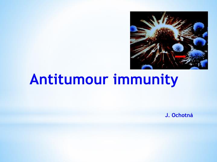
Antitumour immunity J. Ochotná. Malignant transformation Failure of cell division regulation and regulation of cells " social " behavior The uncontrollable proliferation , dissemination to other tissues Mutations in protoonkogenes and antionkogenes. Tumour antigens

E N D
Antitumourimmunity J. Ochotná
Malignanttransformation Failureofcell divisionregulation and regulationofcells "social" behavior Theuncontrollableproliferation, dissemination to othertissues Mutations in protoonkogenes and antionkogenes
Tumour antigens Tumour specific antigens (TSA) a) complexes of MHCgp I with abnormal fragments of cellular proteins chemically induced tumors leukemia with chromosomal translocation b) complexes of MHCgp with fragments of oncogenic viruses proteins tumors caused by viruses (EBV, SV40, polyomavirus) c) abnormal forms of glycoproteins Sialylation of surface proteins of tumor cells d) idiotypes of myeloma and lymphoma clonotyping TCR and BCR
Tumourassociatedantigens (TAA) • expressedalsoon normalcells • differencesin quantity, time and localofexpression • auxiliarydiagnosticmarkers a) onkofetalantigens • on normalembryoniccells and some tumor cells • -fetoprotein (AFP) - hepatom • canceroembryonalantigen (CEA) - coloncancer b)melanomaantigens • MAGE-1, Melan-A
c) antigen HER2/neu receptor forepithelialgrowthfactor mammarycarcinoma d) EPCAM epithelial cell adhesionmolecule metastases e) differentiationantigensofleukemiccells present on normalcellsofleukocyteslinage CALLA -acutelymphoblasticleukemia (CD10, pre-B cells)
Anti-tumor immunemechanisms Hypothesisofimmunecontrol tumor cellsnormallyarise in tissues and are eliminated by Tlymphocytes
Regulatory T cellspreventsremovalofcancercells and thuscontribute to thedevelopmentofthe tumor.
DC are necessary for activation of antigen specific mechanisms • cancer-associated antigens are processed by DC and presented to T lymphocytes in complexes with HLA I. and II. • predominance of TH1 (IFN TNF); IFNg support DC maturation • tumor cells are eliminated byTC • TH2 activation → support B lymphocytes→ tumor specific antibodies (involved in the ADCC) • tumor cells are also destroyed by NK cells (low MHC gpI expression on tumor cells) • interferons - antiproliferative, cytotoxic effect on tumor cells - INFg - DC maturation
Mechanismsof tumor resistance high variability of tumor cells lowexpressionof tumor antigens sialylation tumor cells do not providecostimulatorysignals → T lymphocyteanergy someantitumorsubstanceshave a stimulatingeffect on tumor productionoffactorsinactivating T lymphocytes expressionofFasL → T lymphocyteapoptosis inhibitionofthedendriticcell functions and durability (NO, IL-10, TGF-
Transplantation = transfer of tissue or organ • autologous - donor = recipient • syngeneic - genetically identical donor and recipient (identical twins) • allogeneic - genetically nonidentical donor of the same species • xenogenic - the donor of other species • implant - artificial tissue compensation
Allogeneictransplantation • differencesin donor-recipient MHC gp and secondaryhistocompatibilityAg • alloreactivityof T lymphocytes - the risk ofrejection and graft-versus-host • direct recognitionofalloantigens – recipient T lymphocytesrecognizethedifferent MHC gp and non-MHC molecules on donor APC • indirectrecognitionofalloantigens - APC absorbdifferent MHC gpfromdonor cells and presentthefragments to T lymphocytes • CD8+ T cellsrecognize MHC gp I. • CD4+ T cellsrecognize MHC gp II.
Tests prior to transplantation ABO Compatibility -risk of hyperacute or accelerated rejection = formation of Ab against A or B Ag on graft vascular endothelium) HLA typing (determination of MHC gp alelic forms) phenotyping and genotyping by PCR Cross-match - lymfocytotoxic test - testing preformed Ab (after blood transfusions, transplantation, repeated childbirth) Mixed lymphocyte reaction - test for T lymphocytes alloreactivity
HLA typing a) phenotyping: evaluation of HLA molecules using typing serums Typing antiserums = alloantiserums of multipar (created cytotoxic Ab against paternal HLA Ag of their children), serum of patients after repeated blood transfusions, monoclonal Ab
b) genotyping: PCR – SSP PCR – SSO PCR – SBT
Cross-match test • determination of preformed antibodies • recipient serum + donor lymphocytes + rabbit complement → if cytotoxic Ab against donor HLA Ag are present in recipient serum (called alloantibodies = Ab activating complement) → lysis of donor lymphocytes. Visualization of dye penetration into lysis cells. • positive test = the presence of preformed Ab → risk of hyperacute rejection! → contraindication to transplantation
Mixed lymphocyte reaction (MRL) • determination of T lymphocyte alloreactivity • mixed donor and recipient lymphocytes → T lymphocytes after recognition allogeneic MHC gp activate and proliferate • the total proliferation of lymphocytes is measured by adding [3H]-thymidine to the culture medium and monitoring its integration to DNA of new cells
One-way MRL • determinationof recipient T lymphocytesreactivityagainstdonor cells • donor cellstreatedwithchemotherapyorirradiated lose theabilityofproliferation
Rejection Factors: The genetic difference between donor and recipient, especially in the genes coding for MHC gp (HLA) Type of tissue / organ - the strongest reactions against vascularized tissues containing much APC (skin) The activity of the immune system of the recipient - the immunodeficiency recipient has a smaller rejection reaction; immunosuppressive therapy after transplantation – suppression of rejection Status transplanted organ - the length of ischemia, the method of preservation, traumatization of organ at collection
Hyperacute rejection • minutes to hours after transplantation • antibodies immune response • mechanism: • in recipients blood are present before transplantation preformed or natural Ab (IgM anti-carbohydrate Ag) → Ab + Ag of graft (MHC gp or endothelial Ag) → graft damage by activated complement (lysis of cells) • the graft endothelium: activation of coagulation factors and platelets, formation thrombi, accumulation of neutrophil granulocytes • prevention: • negat. cross match before transplantation, ABO compatibility
Accelerated rejection Hyperacute rejection
Accelerated rejection • 3 to 5 days after transplantation • caused by antibodies that don‘t activate complement • cytotoxic and inflammatory responses activated by antibodies which bind to Fc-receptors on phagocytes and NK cells • prevention: • negative cross match before transplantation, ABO compatibility
Acute rejection • days to weeks after the transplantation or after a lack of immunosuppressive treatment • cell-mediated immune response • mechanism: • recipient TH1 and TC cells response against Ag of graft tissue • infiltration of lymphocytes, mononuclears, granulocytes around smallvessels → destruction of transplant tissue
Chronic rejection • occures after 2 months from transplantation • the most common cause of graft failure • mechanism is not fully understood: • non-immunological factors (tissue ischemia) and TH2 responses with production alloantibodies, pathogenetic role of cytokines and growth factors (TGF β) • replacement of functional tissue by fibrous tissue, endothelial damage →impaired perfusion of graft → gradual loss of its function dominating findings: vascular damage
hematopoetic stem cell collection • myeloablation • transplantation • engraftment • rejection • graft versus host disease Bone marrow transplantation
Graft-versus-host disease (GVHD) • after bone marrowtransplantation • GVHD alsoafterbloodtransfusion to immunodeficiencyrecipients • T-lymphocytes in thegraft bone marrowrecognize recipient tissueAg as foreign (alloreactivity)
Acute GVHD • days to weeksafter stem cellstransplantation • damageof liver, skin and intestinalmucosa • Prevention: appropriate donor selection, T lymphocytesremovalfromthegraft and effectiveimmunosuppression
Chronic GVHD • months to yearsaftertransplantation • TH2 lymphocytesinfiltrationoftissues and organs, productionofalloantibodies and productionofcytokines → fibrosis • processlikeautoimmunedisease: vasculitis, scleroderma, sicca-syndrome • chronicinflammationofbloodvessels, skin, internalorgans and glands, whichleads to fibrosis, bloodcirculationdisorders and lossoffunction
Graft versus leukemiaeffect (GVL) • donor T lymphocytesreactagainstresidualleukemickcellsof recipient • mechanismisconsistentwithacute GVHD • associatedwith a certaindegreeof GVHD (adversereactions)
Classification by Coombs and Gell Immunopathological reactions: immune response, which caused damage to the body (secondary consequence of defense responses against pathogens, inappropriate responses to harmless antigens, autoimmunity) IV types of immunopathological reactions: Type I reaction - response based on IgE antibodies Type II reaction - response based on IgG and IgMantibodies Type III reaction - response based on the formation of immune complexes Type IV reaction - cell-mediated response
Immunopathological reaction based on IgG and IgM antibodies (reaction type II) Cytotoxic antibodies IgG and IgM: • complement activation • ADCC • binding to phagocytes and NK cells Fc receptors Haemolytic reactions after transfusion of ABO incompatible blood: Binding of antibodies to antigens of erythrocytes → activation of the classical way of complement → cell lysis Hemolytic disease of newborns: Caused by antibodies against RhD antigen
Autoimmune diseases: • organ-specific cytotoxic antibodies (antibodies against erythrocytes, neutrophils, thrombocytes, glomerular basement membrane ...) • blocking or stimulating antibodies Graves - Basedow disease - stimulating antibodies against the TSH receptor Myasthenia gravis - blocking of acetylcholin receptor→ blocking of neuromuscular transmission Pernicious anemia - blocking of vitamin B12 absorption Antiphospholipid syndrome - antibodies against fosfolipids Fertility disorders - antibodies against sperms or oocytes
Immunopathologicalreactionsbased on immunecomplexformation (reaction type III) • caused by IgGantibodies → bind to antigen → creationofimmunecomplexes • immunocomplexes - bind to Fcreceptors on phagocyte - activatecomplement • immunecomplexes (depending on thequantity and structure)are eliminated by phagocytesorstored in tissues • pathologicalimmunocomplexes response ariseswhenis a largedose of antigen, or antigen in the body remains • immunecomplexes are deposited in thekidneys (glomerulonephritis), on theendothelialcellssurface (vasculitis) and in synovialjoints(arthritis)
Serumsickness • aftertherapeuticapplicationofxenogeneicserum (antiserum to snakevenom) • creationofimmunecomplexes and theirstoragein thevesselwallsofdifferentorgans • clinicalmanifestations: urticaria, arthralgia, myalgia • Systemic lupus erythematosus • antibodiesagainstnuclearantigens, ANA, anti-dsDNA • Farmer'slung • IgGantibodiesagainstinhaledantigens (molds, pollens) Poststreptococcalglomerulonephritis
Immunopathological delayed-type reaction (reaction type IV) • delayed-type hypersensitivity (DTH) • local reaction caused by TH1 cells and monocytes / macrophages (physiologically – elimination of macrophage intracellular parasites) • antigen immunization → formation of antigen specific TH1 cells (and memory cells) • 12-48 hours after next contact with antigen arise local reaction – granuloma (TH1and macrophage infiltration) • Tuberculin reaction Tissue damage in tuberculosis and leprosy Sarcoidosis
similar to DTH reaction • TH1 cells activate CD8 + T lymphocytes viral rashes viral hepatitis acute rejection of transplanted organ some autoimmune thyroiditis contact dermatitis Subtype IV - Cellularcytotoxic response(Tcactivation)
Contact dermatitis • is a localizedrashorirritationofthe skin causedby contactwith alergen (nickel , chromium, ingredients in cosmeticproducts , plant allergens and other) • thefirstissenzitization • appears in 24 – 48 hoursafter second contactwith alergen • diagnosis : patch test
patch testis a methodused to determineif a specific substance causesallergicinflamationofthe skin • Allergens are applied to specialhypoallergenicpatch on theback skin • Results are evaluatedafter48 and 72 hours • In positive reactionappearseczema Patch test