Download
1 / 45
590 likes | 1.78k Vues
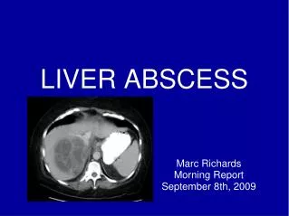
LIVER ABSCESS. Occurs when bacteria/protozoa destroy hepatic tissue, produces a cavity which fills up with infective organisms, liquefied cells & leucocytes. Necrotic tissue then falls off the cavity from rest of the liver. Pyogenic abscess Amoebic abscess. Pyogenic abscess.

E N D
Occurs when bacteria/protozoa destroy hepatic tissue, produces a cavity which fills up with infective organisms, liquefied cells & leucocytes. Necrotic tissue then falls off the cavity from rest of the liver.
Pyogenic abscess • Amoebic abscess
Pyogenic abscess • Male preponderance • Average age – between 43 & 60 years • Infective organisms invade liver directly after liver wound or spread from lungs, skin or other organs by hepatic artery, portal vein, biliary tract
Origin & causes • Biliary tract Underlying biliary diseases is the most common cause; a) Septic cholangitis b) Biliary stenting c) Sclerosing cholangitis d) Cholangio carcinoma
2) Portal vein Portal pyaemia following pelvic / GI infection that leads to Portal Pylephlebitis or septic emboli a) Appendicitis b) Diverticulitis c) Perforated ulcer b) Pancreatitis
3) Hepatic artery a) Bacterial endocarditis 4) Trauma a) Penetrating wound b) Blunt trauma
5) Direct a) Perforated peptic ulcer b) Subphrenic abscess c) Adjacent abscess- Perinephric abscess
6) Miscellaneous Obscure in 5 % cases Other unusual causes: a) Cysts- Including Polycystic liver disease) b) Intrahepatic malignancy c) Hydatid cyst
Bacteriology • Majority derived from GI tract in >75% cases • Aerobic: a) E.coli – most common cause b)Klebsiella pneumoniae c)Others: Pseudomonas aerogenosa, Morganella morganii, Serratia marsecens, etc.
Anaerobic: a) Bacteroides fragilis (most common) b) Others: Fusobacterium spp., anaerobic Streptococci, Clostridium spp., Actinomyces spp.
Based on size & distribution of focal sites; a) Macroscopic abscess b) Microscopic abscess
Clinical features • Symptoms: Fever Abdominal pain Chills Anorexia Weight loss Nausea, Vomiting Right shoulder pain / irritable cough
Signs: Hepatomegaly Tenderness Rebound tenderness Jaundice (late)
Histology • Portal zone infection & surrounding hepatocytes, infiltrated with polymorph
Investigations 1) Routine: a) Hb-anemia b) WBC-luecocytosis c) Blood culture – organisms present 2) Liver Function test: a) Elevated serum alkaline phosphatase (most reliable); b) Elevated serum bilirubin (50%) c) Elevated aminotransferases (48%) d) Hypoalbuminemia (33%)
3) Chest X-Ray: a) Elevation of right hemidiaphragm b) Right basilar infiltrate 4) USG: Differentiate as a round or oval area- hypoechoic fluid filled area 5) CT Scan: Cluster sign- seen when multiple small abscess aggregate, which suggests beginning of coalescence to single abscess
6) MRI: Raised lesion with sharp borders 7) Aspiration of material: Diagnostic & Therapeutic
Treatment Start with empiric antibiotics: Ampicillin Metronidazole Gentamicin Specific antibiotics (Depending on cultures) 6-16 weeks If persisting Percutaneous drainage (under USG/CT guidance) Surgical Drainage
Indications for surgical drainage: a) Abscess with intra-abdominal pathology requiring surgery b) Ascitis c) Multiple large abscesses d) Abscess which cannot be drained percutaneously
Amoebic Abscess • Commonest extra-intestinal presentation of amoebiasis • Common in alcoholics • Caused by Entamoeba histolytica • Entry by faeco-oral route
Pathology • Amoeba multiply-block in intrahepatic portal radicles-focal infarction of liver cells- proteolytic enzymes released- destiny liver parenchyma
Site: Right lobe of liver, supraanteriorly, just below the diaphragm Large necrotic area which is liquefied into thick reddish-brown pus (Anchovy sauce pus) due to liquefied necrosis, thrombosis of blood vessels, lysis of liver cells
Histology Necrotic area containing degenerated liver cells, leucocytes, RBCs, connective tissue strands, debris & amoeba
Clinical features • Symptoms: High grade fever with rigor Weight loss Upper quadrant pain ( Initially dull aching, later on stabbing) Jaundice (not common)
Signs: Hepatomegaly (tender) Consolidation in right lower zone of lungs Pleurisy
Complications 1) Rupture into lung/pleura a) Empyema b) Hepatobronchial fistula c) Pulmonary abscess d) Pneumonitis e) Pleural effusion
2) Rupture into pericardium 3) Intraperitoneal rupture 4) Rupture into portal vein (rare) 5) Secondary infection
Investigations • Routine: Leucocytosis Anemia 2) Liver function test: Increased Alkaline Phosphatase Increased Transaminase 3) Stool examination: cysts/ trophozoites
4) Aspiration: Anchovy sauce pus 5) Chest X-Ray Raised fixed diaphragm Right lateral abscess 6) USG (most useful) : Round lesion 7) CT : Irregular edge 8) Serology: ELISA
Treatment • Metronidazole 750mg orally/i.v. 3 daily x 4 days If response, continue for 10 days; followed by luminal agents: Iodoquinol 650mg 3 X 20 days Paramomycin 500mg 3 X 10 days
If no response, Dihydroemetine 1.5mg/Kg i.m. 4 X 5 days + Chloroquine phosphate 600mg base/day orally 4 X 2 days, then 300mg base/day orally 4 times If no response to medical treatment: Percutaneous drainage