Download
1 / 57
620 likes | 802 Vues
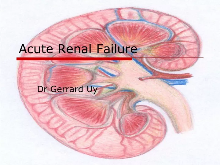
Acute Renal Failure. Dr Gerrard Uy. Overview. Definitions Classification and causes Presentation Treatment. Definition Acute Renal failure (ARF). Inability of kidney to maintain homeostasis leading to a buildup of nitrogenous wastes

E N D
Acute Renal Failure Dr Gerrard Uy
Overview • Definitions • Classification and causes • Presentation • Treatment
Definition Acute Renal failure (ARF) • Inability of kidney to maintain homeostasis leading to a buildup of nitrogenous wastes • Different to renal insufficiency where kidney function is deranged but can still support life • Characterized by a rapid decline in glomerular filtration rate (GFR)
ARF • Occurs over hours/days • Usually asymptomatic • Lab definition • Increase in baseline creatinine of more than 50% • Decrease in creatinine clearance of more than 50% • Deterioration in renal function requiring dialysis
ARF definitions • Anuria – no urine output or less than 100mls/24 hours • Oliguria - <500mls urine output/24 hours or <20mls/hour • Polyuria - >2.5L/24 hours
ARF • Pre renal (functional) • Renal-intrinsic (structural) • Post renal (obstruction)
ARF Pirouz Daeihagh, M.D.Internal medicine/Nephrology Wake Forest University School of Medicine. Downloaded 4.6.09
Causes of ARF • Pre-renal: Inadequate perfusion • check volume status • Renal: ARF despite perfusion & excretion • check urinalysis, FBC & autoimmune screen • Post-renal: Blocked outflow • check bladder, catheter & ultrasound
ARF Pre renal • Most common form of ARF • Decreased renal perfusion without cellular injury • 70% of community acquired cases • 40% hospital acquired cases
ARF Pre renal • Hepatorenal syndrome • Unique form of prerenal ARF • Frequently complicates advanced cirrhosis • Kidneys are structurally normal but fail due to splanchnic vasodilation and arteriovenous shunting • 2 types: • Type 1 HRS – more aggressive • Type 2 HRS
ARF Intrinsic • Acute tubular necrosis (ATN) • Ischaemia • Toxin • Tubular factors • Acute interstitial Necrosis (AIN) • Inflammation • oedema • Glomerulonephritis (GN) • Damage to filtering mechanisms • Multiple causes as per previous presentation
ARF Post renal • Post renal obstruction • Obstruction to the urinary outflow tract • Bladder neck obstruction – most common cause of post renal ARF • Prostatic hypertrophy • Blocked catheter • Malignancy
Prerenal Failure 1 • Often rapidly reversible if we can identify this early. • The elderly at high risk because of their predisposition to hypovolemia and renal atherosclerotic disease. • This is by definition rapidly reversible upon the restoration of renal blood flow and glomerular perfusion pressure. • THE KIDNEYS ARE NORMAL. • This will accompany any disease that involves hypovolemia, low cardiac output, systemic dilation, or selective intrarenal vasoconstriction. ARF Anthony Mato MD Downloaded 5.8.09
Differential Diagnosis 2 • Hypovolemia • GI loss: Nausea, vomiting, diarrhea (hyponatraemia) • Renal loss: diuresis, hypo adrenalism, osmotic diuresis (DM) • Sequestration: pancreatitis, peritonitis,trauma, low albumin (third spacing). • Hemorrhage, burns, dehydration (intravascular loss). ARF Anthony Mato MD Downloaded 5.8.09
Differential Diagnosis 3 • Renal vasoconstriction: hypercalcaemia, adrenaline/noradrenaline, cyclosporin, tacrolimus, amphotericin B. • Systemic vasodilation: sepsis, medications, anesthesia, anaphylaxis. • Cirrhosis with ascites • Hepato-renal syndrome • Impairment of autoregulation: NSAIDs, ACE, ARBs. • Hyperviscosity syndromes: Multiple Myeloma, Polycyaemia rubra vera
Differential Diagnosis 4 • Low CO • Myocardial diseases • Valvular heart disease • Pericardial disease • Tamponade • Pulmonary artery hypertension • Pulmonary Embolus • Positive pressure mechanical ventilation
Glomerular blood flow Compensatory Dilators: Prostacyclin, NO Blocker: NSAID Glomerular Capillaries & Mesangium Afferent arteriole Efferent art Blocker: ACE-I Compensatory Constrictor: Angiotensin II Constrictors: endothelin, catecholamines, thromboxane
Acute Tubular Necrosis (ATN)Classification • Ischemic • Nephrotoxic
Acute Renal FailureNephrotoxic ATN • Endogenous Toxins • Heme pigments (myoglobin, hemoglobin) • Myeloma light chains • Exogenous Toxins • Antibiotics (e.g., aminoglycosides, amphotericin B) • Radiocontrast agents • Heavy metals (e.g., cis-platinum, mercury) • Poisons (e.g., ethylene glycol)
Acute Interstitial NephritisCauses • Allergic interstitial nephritis • Drugs • Infections • Bacterial • Viral • Sarcoidosis
Allergic Interstitial Nephritis(AIN)Clinical Characteristics • Fever • Rash • Arthralgias • Eosinophilia • Urinalysis • Microscopic hematuria • Sterile pyuria • Eosinophiluria
Contrast-Induced ARFPrevalence • Less than 1% in patients with normal renal function • Increases significantly with renal insufficiency
Contrast-Induced ARFRisk Factors • Renal insufficiency • Diabetes mellitus • Multiple myeloma • High osmolar (ionic) contrast media • Contrast medium volume
Contrast-induced ARFClinical Characteristics • Onset - 24 to 48 hrs after exposure • Duration - 5 to 7 days • Non-oliguric (majority) • Dialysis - rarely needed • Urinary sediment - variable • Low fractional excretion of Na
Pre-Procedure Prophylaxis 1. IV Fluid (N/S) 1-1.5 ml/kg/hour x12 hours prior to procedure and 6-12 hours after 2. Mucomyst (N-acetylcysteine) Free radical scavenger; prevents oxidative tissue damage 600mg po bd x 4 doses (2 before procedure, 2 after) 3. Bicarbonate (JAMA 2004) Alkalinizing urine should reduce renal medullary damage 5% dextrose with 3 amps HCO3; bolus 3.5 mL/kg 1 hour preprocedure, then 1mL/kg/hour for 6 hours postprocedure 4. Possibly helpful? Fenoldopam, Dopamine 5. Not helpful! Diuretics, Mannitol
Contrast-induced ARFProphylactic Strategies • Use I.V. contrast only when necessary • Hydration • Minimize contrast volume • Low-osmolar (nonionic) contrast media • N-acetylcysteine, fenoldopam
ARF Post-renal Causes 1 • Intra-renal Obstruction • Acute uric acid nephropathy • Drugs (e.g., acyclovir) • Extra-renal Obstruction • Renal pelvis or ureter (e.g., stones, clots, tumors, papillary necrosis, retroperitoneal fibrosis) • Bladder (e.g., BPH, neuropathic bladder) • Urethra (e.g., stricture)
Acute Renal FailureDiagnostic Tools • Urinary sediment • Urinary indices • Urine volume • Urine electrolytes • Radiologic studies
Urinary Sediment (1) • Bland • Pre-renal azotaemia • Urinary outlet obstruction
Urinary Sediment (2) • RBC casts or dysmorphic RBCs • Acute glomerulonephritis • Small vessel vasculitis
Red Blood Cells Monomorphic Dysmorphic
Urinary Sediment (3) • WBC Cells and WBC Casts • Acute interstitial nephritis • Acute pyelonephritis
Urinary Sediment (4) • Renal Tubular Epithelial (RTE) cells, RTE cell casts, pigmented granular (“muddy brown”) casts • Acute tubular necrosis
Acute Renal FailureUrine Volume (1) • Anuria (< 100 ml/24h) • Acute bilateral arterial or venous occlusion • Bilateral cortical necrosis • Acute necrotizing glomerulonephritis • Obstruction (complete) • ATN (very rare)
Acute Renal FailureUrine Volume (2) • Oliguria (<100 ml/24h) • Pre-renal azotemia • ATN • Non-Oliguria (> 500 ml/24h) • ATN • Obstruction (partial)
Clinical Assessment • Thirst and orthostatic dizziness • Orthostatic hypotension • Tachycardia • Reduced JVP • Decreased skin turgor • Dry mucous membranes
ARF-Signs and Symptoms • Weight gain • Peripheral oedema • Hypertension
ARF Signs and Symptoms • Hyperkalemia • Nausea/Vomiting • Pulmonary edema • Ascites • Asterixis • Encephalopathy