Download
1 / 61
610 likes | 628 Vues
ACUTE RENAL FAILURE. DEFINITION:. It is abrupt decrease in renal function which developed over a period of days or weeks and usually accompanied by reduction in urine volume and retention of nitrogenous waste (( eg. Blood urea and createnin )). CAUSES:. 1. Pre renal:

E N D
DEFINITION: • It is abrupt decrease in renal function which developed over a period of days or weeks and usually accompanied by reduction in urine volume and retention of nitrogenous waste (( eg. Blood urea and createnin ))
CAUSES: 1. Pre renal: a. Absolute decrease in effective blood volume eg. Hemorrhage, burns, diarrhea, vomiting. b. Relative decrease in effective blood volume eg. Congestive heart failure, sepsis, anaphylaxis c. Arterial occlusion
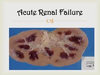
2. Renal: a. Acute tubular necrosis (( ATN )) 1. Ischemic insult ( prolonged hypotension ) 2. Nephrotoxic insult which is either: • Exogenous nephrotoxins eg. Aminoglycosides, amphotericin B, contrast agents. • Endogenous substances: Haemoglobuline, myoglobulin, uric acid, bacterial toxins.
b. Acute interstitial nephritis c. Acute glomerulonephritis
3. Post renal a. Obstructive uropathy b. Bladder neck obstruction.
**Reversible pre renal ARF: • Marked hypotension and signs of poor peripheral perfusion • Postural hypotension(fall > 20/10 mmHg ) • Evidence of the underlying cause but some time concealed blood loss can occur eg GI-bleeding, pelvic fracture
Metabolic acidosis and hyperkalaemia are often present • Significant oliguria with concentrated urine • Elevated blood urea level with no or minimal increase in serum creatinine. • (B.urea/S.creatinine ratio > 40 ). • Low urine sodium < 20 meq/L.
Management: 1. Establish and correct the underlying cause of ARF. 2. If the patient is hypovolaemic, restore the blood volume as rapidly as possible with blood, plasma or normal saline. 3. Vital signs monitoring and central venous pressure monitoring.
4. Correct metabolic acidosis by: a. Restoration of blood volume. b. Isotonic Sodium bicarbonate may be used. * If the treatment given sufficiently early, renal function will improve rapidly but in some cases the treatment is ineffective and renal failure become established.
CLINICAL FEATURES: 1. Features of the underlyingcause eg. Trauma, sepsis 2. Alteration in urine volume usually started as oliguric phase followed by polyuric phase which carry better prognosis and may indicate regeneration and recovery from ATN.
3.GIT symptoms ((early uremic symptoms)) include: anorexia, nausea, vomiting and hiccoughs. 4. Neurological manifestations: drowsiness,apathy,confusion, fit, muscle twitching and comma.
5.Cardiovascularfeatures: : pericarditis and arrhythmias which may be due to uremia itself or as a resut of electrolyte disturbances.
6. Electrolytes and metabolic abnormalities: a. Hyperkalemia: particularly with massive tissue break down or haemolysis, ECG changes ((tinted T-wave, absence of P-wave, wide QRS complex and Sine wave)), elevated serum potassium.
b. Metabolic acidosis. c. Dilutional hyponatermia. d.Hypocalcaemia& Hyperphosphatemia.
7.Increased respiratory rate due to acidosis, pulmonary oedema or respiratory infection. 8. Anaemia may be present due to excessive blood loss or it may indicate acute exacerbation on CRF.
9.Severe infection may complicate ARF due to decreased immunity. 10. Volume overload: bilateral leg oedema, Pulmonary oedema, Pericardial effusion.
** Clinical approach to the diagnosis of ARF: The clinical evaluation of ARF is achieved by answering the following five questions :
Is it ARF or an acute on chronic renal failure? Is there renal tract obstruction? Is there reduction in effective ECF volume? Has there been a major vascular occlusion? Is there parenchymal renal disease other than ATN?
A complete history, physical examination and chart review, as well as a careful urinalysis, a renal ultrasound, and a few routine blood tests should be performed in all patients with ARF .
This approach will reveal the likely cause of ARF in most patients. In a few carefully selected patient ,additional ,selected special investigations-imaging, serology, renal biopsy-may be necessary to establish the cause of renal dysfunction.
History The diagnosis of prerenal failure is often facilitated by careful evaluation of the patient fluid balance during the few days preceding the onset of ARF.
H/O vomiting or diarrhea provides useful clues to the source of loss of ECF volume. Symptoms of dry cough,orthopnea, or ankle swelling may indicate the possibility of early volume overload.
Frequency, urgency, and hesitancy are important symptoms in patient with bladder dysfunction or bladder neck obstruction. H/O ingestion of a nephrotoxic drugs
Systemic symptoms such as fever, malaise or fatigue, and joint pain or skin rash raise the possibility of ARF caused by acute GN associated with SBE, vasculitis or connective tissue disease like SLE H/O previous renal disease or hypertension may point to pre-existing chronic renal insufficiency.
Chart review Since most ARF occurs in patients in hospital,the hospital record represent an extremely an important source of information regarding the possible cause. A chart review should be considered as an extension of the history, daily record of vital signs, changes in body weight and records of fluid intake and out put. The record of all drugs taken by the patient must be examined for potentially nephrotoxic drugs.
PHYSICAL EXAMINATION *Loss of skin turgor and postural hypotension when fluid loss exceed 5-10% of ECF volume. *Supine hypotension when the fluid loss exceed 20% of ECF volume. *Raised JVP ,pedal edema and pulmonary crackles indicate cardiac failure.
Skin: *Jaundice and other evidence of acute or chronic liver disease *Maculopapularskin rash may indicate drug induced AIN or acute GN. *Malar rash or photosensitivity in SLE *Livedoreticularisassociated with cryoglobulinemia or atheroembolic disease
*Palpable purpura commonly caused by cutaneous vasculitis or atheroembolic disease *Nonpalpable purpura may suggests the presence of thrombocytopenia associated with SLE or thrombotic microangiopathy.,
Eye examination *Jaundice, hypertensive retinopathy, Roth spots (endocarditis)or cholesterol emboli (atheroembolic disease)
Abdominal examination: *Ascites may indicate chronic liver disease or congestive cardiac failure *Bruits over the anterior abdomen or confirmed to the renal angles
*Any evidence of bladder neck obstruction *Rectal or pelvic examination *The presence of large postvoid residual (more than 200-300 ml) is strongly suggestive of functionally significant urinary retention.
Urine volume Patient with prerenal azotemia almost always have oliguria, but non oligureic ARF seen in : 1. diabetes insipidus 2. sodium wasting disease such as adrenal insufficiency 3. hyperglycemia
4. malnutrition 5. chroniclly ill patient 6. urinary tract obstruction ( unilateral or partial ) 7. contrast induced ARF 8. aminoglycosid induced ARF
*Anuria is unusual in patient with ATN but it is more common with rapidly progressive GN, acute interstitial nephritis or vascular catastrophes such as renal artery embolism or renal vein thrombosis .
If patient thought to have ATN have persistent anuria lasting more than 24 -48 hours, the other causes of ARF must be reconsidered.
Blood urea nitrogen *The clearance of urea is always less than the GFR. This is because of the back diffusion of urea which is inversely related to the rate of urine flow and is enhanced by vasopressin. *BUN rises more rapidly than the creatinine in patient with Prerenal failure
The ratio BUN : Cr is normally 15:1 (mg:mg) (60:1 mmol:mmol) High ratio > 20:1 caused by: 1. Prerenal failure 2. GI bleeding 3. increased protein intake
4. catabolic state 5. infusion of amino acids 6. corticosteroid therapy 7. tetracycllines
Low ratio < 5-10 :1 caused by: 1. severe liver disease2. malnutrition3. rhabdomyolysis4. cimetidine5. trimethoprim6. cephalosporins
SERUM CREATININE *As renal function deteriorates ,the Cr becomes progressively less reliable as an index of GFR.. *BUN rarely exceeds 30mg/dl.and serum Cr > 2 mg/dl.unless there is concurrent underlying chronic renal insufficiency.
OTHER SERUM CHEMISTRY: *They are usually unhelpful in the diagnostic process. Hyperkalemia, hyperuricemia, hypocalcemia and hyperphosphatemia are all common in ARF of any cause also they are not useful in distinguishing ARF from CRF
GUE may shows evidence of underlying renal pathology eg. GN, Interstitial nephritis or evidence of underlying cause eg DM, Multiple myeloma
Hematological indices -the hematocrit is not useful discriminate between ARF and CRF. -High WBC count may point to systemic infection, lymphoma and leukemia - Eosinophelia may suggest a drug induced ATN or atheroembolic disease - Thrombocytopenia suggest HUS,TTP, SLE, myeloma, sepsis, and DIC.
RENAL ULTRASOUND - It is a key investigation for the diagnosis of obstruction or CRF. - It is not a reliable method for identifying the anatomical site of obstruction - Even if the kidneys are reduced in size , the possibility of Prerenal ARF or ATN superimposed on CRF must always be considered.