Download
1 / 62
620 likes | 622 Vues
Learn about the essential tasks performed by medical assistants in the microbiology laboratory, including specimen collection and testing, culture preparation, and identification of organisms. Discover the importance of sensitivity testing and techniques to avoid laboratory errors.

E N D
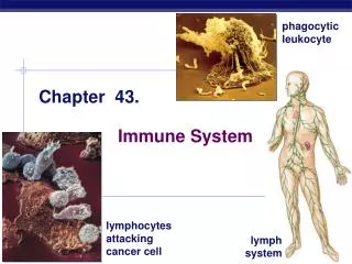
Chapter 43 Basic Microbiology
The Medical Assistant’s Role in the Microbiology Laboratory • Obtain and test specimens • Prepare slides and cultures • Allow cultures to grow at least 12 hours before examining for identification • Sensitivity identifies which antibiotic(s) will kill microorganism causing infection
The Medical Assistant’s Role in the Microbiology Laboratory • Normal flora: natural bacteria • Pathogen: disease causative microorganism • Use technique to avoid laboratory error • Use sterile supplies • Deliver culture to laboratory in reasonable amount of time
The Medical Assistant’s Role in the Microbiology Laboratory • Identification of organisms done successfully within 24 to 72 hours • Many test kits available
Microbiology • Classification • Taxonomy deals with classification of living organisms • Kingdoms: plants, animals, protists • Lower protists, or prokaryotes (blue-green algae and bacteria) • Higher protists, or eukaryotes (protozoa, algae, fungi)
Microbiology • Nomenclature • System for naming bacteria • Genus • First name; capitalized • Species • Second name; not capitalized
Microbiology • Nomenclature • Bacteriologists and microbiologists • Parasitology • Virology • Mycology • Reference laboratory • Report certain types of bacteria and yeasts to Department of Public Health
Microbiology • Cell structure • DNA (deoxyribonucleic acid) • Lower protist Basic bacterial cell >>
Equipment • Autoclave • Used to sterilize equipment • Many laboratories no longer use autoclaves because of use of presterilized and disposable equipment
Equipment • Microscope • Used to view organisms that cannot be seen with naked eye on prepared slide • Delicate instrument • Cared for properly as stated by manufacturer
Equipment • Safety hood • Aerosols can be released into air when culturing • Potentially dangerous if inhaled • Use of hood mandatory when performing culture on specimen with potential aerosol • Used to minimize odors
Equipment • Incubator • Constant temperature of 35–37°C • Grows aerobic or anaerobic organisms • Temperature requirements must be met for adequate growth
Equipment • Anaerobic equipment • Absence of oxygen to grow anaerobic bacteria • Use of candle jar • Gas pack jar • Specimens sent to reference laboratories • Gram stain used to observe gross morphological features of bacteria
Equipment • Inoculating equipment • Loop used to inoculate organisms • Needle used when performing stab culture Inoculating loop>> Inoculating needle>>
Equipment • Incinerator • Quickest method of sterilization • Electrical incinerator or Bunsen burner • Media • Host of substances • Used to foster growth of bacteria
Equipment • Refrigerator • Used to store materials • Temperature of 2–8°C • Never store food or drink with specimens, kits, media
Safety When HandlingMicrobiology Specimens • Personal protective equipment • PPE worn at all times • Laboratory coat or apron, safety goggles, gloves • May need to work behind shield or use safety hood • Never eat, smoke, drink, or put objects into mouth • Do not touch contact lenses or apply makeup • Wash hands
Safety When HandlingMicrobiology Specimens • Work area • Use strong germicide before and after daily use or immediately after spills • Dust-free and clean at all times • Uncluttered • Avoid body burns or files
Safety When HandlingMicrobiology Specimens • Specimen handling • Look for leaks and contamination on containers • Wear gloves • Use appropriate container • Handle all specimens as if contaminated
Safety When HandlingMicrobiology Specimens • Disposal of waste and spills • Separation of biohazardous wastes (red bags) • Disinfect spills with 5% phenol or 10% bleach solution <<Biohazard symbol
Quality Control • All equipment with temperature controls monitored daily • Microscopes cleaned and kept dust-free • Before use, expiration date checked on all testing kits
Quality Control • Media of all types should not be used past shelf life and should be stored at proper temperatures • Check specific list of bacteria to use on various media to test for growth • Laboratory manual updated periodically
Quality Control • All chemicals or reagents with Material Safety Data Sheets (MSDS) should be available to reference • Document all quality control testing in proper laboratory logs
Collection Procedures • Check to see if culture was: • Collected properly • Delivered within a reasonable period of time • Collected in sufficient quantity
Collection Procedures • All specimens taken from site of infection • Place in appropriate container • Deliver to laboratory • Rejecting specimens
Collection Procedures • Factors determining successful isolation of causative pathogens • Proper collection from infection site • Collection of specimen during infection period • Sufficient amount of specimen • Appropriate specimen container • Appropriate transport medium
Collection Procedures • Factors determining successful isolation of causative pathogens • Specimen labeled properly • Specimen delivered to laboratory in minimal amount of time • Specimen collected before administration of antibiotics • Specimen inoculated onto proper media and placed in correct atmosphere to ensure growth
Specific Collection Requirements for Cultures • Urine • Obtaining clean-catch specimen • Use of catheterization • Nose • Nasal-pharyngeal swab collects specimen • Place swab in sterile tube for transport to laboratory
Specific Collection Requirementsfor Cultures • Throat • Specimens taken using culturette • Use sterile tongue depressor to hold patient’s tongue down • Avoid swabbing sides of mouth and tongue • Take specimen directly from affected area
Specific Collection Requirements for Cultures • Wound • Use of sterile needle or swab to aspirate pus-filled fluid from wound • Use of anaerobic transport medium
Specific Collection Requirements for Cultures • Sputum • Patient coughs deeply and expectorates into sterile container • Should be morning specimen • Use of special sterile container
Specific Collection Requirements for Cultures • Stool • Ova and parasites • Bacterial cultures • Keep at between room and body temperature • Non-sterile containers
Specific Collection Requirements for Cultures • Cerebrospinal fluid (CSF) • Lumbar puncture • Fluid dispersed in several departments of clinical laboratory • Use of incubator • Refrigeration can kill meningitis-causing bacteria • STAT order for processing
Specific Collection Requirements for Cultures • Blood • Development of septicemia • Cultures collected by same means as regular blood collection • Variety of collection devices available
Foodborne Illnesses • Bacterial infections and parasites • 48 million cases of “food poisoning” each year contracted from: • Raw or undercooked meats and seafood • Contaminated food handlers • Unwashed produce
Foodborne Illnesses • Symptoms: mild to severe diarrhea, nausea, vomiting, abdominal pain, fever, headaches, flu-like symptoms • Diagnostics: stool cultures, blood tests, following the symptoms • Treatments: antibacterial medications and treating symptoms
Bacterial Shapes Bacilli Cocci Spirilla
Microscopic Examination of Bacteria • Dyes (stains) • Derived from coal tar • Basic dyes carry a positive ion • Acidic dyes carry a negative ion
Microscopic Examination of Bacteria • Simple stain • Uses single stain on fixed slide for given period of time • Shows arrangement and structure of bacterial cell • Takes no more than 3 minutes to stain • Does not give much information
Microscopic Examination of Bacteria • Differential stain • Stain result varies • Common differential stain is Gram stain • Developed in 1884 by Dr. Hans Christian Gram • Differentiates bacteria by Gram stain ability of being negative or positive • Retain or lose color through decolorization • Use of gentian or crystal violet reagents
Microscopic Examination of Bacteria • Differential stain • Identifies Gram-positive and Gram-negative bacteria • Staphylococcus • Streptococcus • E. coli • Proteus • Morphological arrangement, shape, and Gram-stain characteristic help identify bacteria
Microscopic Examination of Bacteria • Acid-fast stain • Specific stain • Allows microscopic examination of acid-fast mycobacteria • Use of heat or powerful dye • Ziehl-Neelsen stain • Kinyoun stain
Microscopic Examination of Bacteria • Special techniques • Used when flagella, spore, capsule, or nuclei of cells present • Organisms in living state, without staining • Wet-mount preparation • Hanging drop preparation
Microscopic Examination of Bacteria • Potassium hydroxide (KOH) preparation • Used for study of fungi and spores • Fragments of human hair, skin, or nails placed on slide with drop of 10% KOH and coverslip • KOH clears debris
Microscopic Examination of Bacteria • Potassium hydroxide (KOH) preparation • Set slide at room temperature for a half hour before examination for debris settlement • Direct examination of specimens • Use of phase or dark-field microscope • Dispose of properly (live organisms)
Culture Media • Inoculate material on proper culture medium for growth and identification • Reliability of results • Aerobic bacteria grow only in oxygen • Anaerobic bacteria live and grow in absence of oxygen
Culture Media • Transport media for reference laboratory • Media solid, liquid, or semisolid substance • Agar: solid form of media • Semisolid media: made by adding less agar • Liquid media: in broth form stored in tubes
Culture Media • Media nutrients to support growth of bacteria • Vitamins • Sugar • Salt • Minerals • Amino acids • Addition of special products
Culture Media • Media classification • Basic • Differential • Selective • Enriched • Check known organisms for quality control and for contaminants
Microbiology Culture • Inoculating the media • Roll swab onto upper quadrant of agar plate • Use loop to inoculate sputum or liquid • Inoculum spread back and forth in sweeping motion with flamed loop or needle