Download

1 / 29
290 likes | 569 Vues
Alterations in the immune response. Anca Ba cârea , Alexandru Schiopu. Allergic and hypersensitivity disorders. Definition An exaggerated immune response to a foreign agent resulting in injury to the host.

E N D
Alterations in theimmune response Anca Bacârea, Alexandru Schiopu
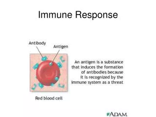
Allergic andhypersensitivitydisorders • Definition • An exaggerated immune response to a foreign agent resulting in injury to the host. • Allergic or hypersensitivity disorders are caused by immune responses to environmental antigens - exogenous and endogenous - that produce inflammation and cause tissue injury. • In the context of an allergic response, these antigens usually are referred to as allergens. • Allergens are any foreign substances capable of inducing an immune response. • Exposure to the allergen can be through inhalation, ingestion, injection, or skin contact.
Classification • Type I, IgE-mediated disorders • Type II, antibody-mediated (cytotoxic) disorders • Type III, immune complex-mediated disorders • Type IV, cell-mediated hypersensitivity
Type I, IgE-Mediated Disorders = Anaphylactic (immediate) hypersensitivity • Mechanism • IgE-mediated—mast cell degranulation • Trigger • binding of an allergen to a specific IgE that is found on the surface of mast cells or basophils • E. g. : Hay fever, asthma, anaphylaxis
Mast cells • Mast cells normally are distributed throughout connective tissue, especially in areas beneath the skin and mucous membranes of the respiratory, gastrointestinal, and genitourinary tracts, and adjacent to blood and lymph vessels - surfaces that are exposed to environmental antigens and parasites. • Mast cells and basophils have granules that contain potent mediators of allergic reactions. • These mediators are preformed in the cell or activated through enzymatic processing.
Mast cells • Sensitization or priming stage • The allergen-specific IgE antibodies attach to receptors on the surface of mast cells and basophils. • Subsequent exposure • The sensitizing allergen binds to the cell associated IgE and triggers a series of events that ultimately lead to degranulation of the sensitized mast cells or basophils, causing release of their allergy-producing mediators.
Type I mediators • The primary (preformed) mediators of allergic reactions include: • Histamine • potent vasodilator that increases the permeability of capillaries and venules • bronchoconstriction • increased secretion of mucus • Acetylcholine • bronchial smooth muscle contraction • dilation of small blood vessels • Adenosine • Chemotactic mediators - cytokines that recruit and activate a variety of inflammatory cells. • Neutral proteases • generate kinins and cleave complement components to produce additional chemotactic and inflammatory mediators
Type I mediators • Secondary mediators - generated from arachidonic acid in the mast cell membrane: • Leukotrienes • Prostaglandins • Their effects are similar to those of histamine and acetylcholine, but delayed and prolonged by comparison. • Platelet-activating factor • Platelet aggregation • Histamine release • Bronchospasm • Chemotactic factor for neutrophils and eosinophils
Presentation of disease • Type I hypersensitivity reactions may present as: • a systemic disorder (anaphylaxis) • localized reaction (atopy)
Systemic anaphylactic reactions • Often result from injected allergens (e.g., penicillin, radiographic contrast dyes, bee or wasp stings). • More rarely, they may result from ingested allergens (seafood, nuts, legumes). • In sensitized individuals, only a small amount of the allergen may be required to produce a reaction.
Systemic anaphylactic reactions - manifestation • Anaphylaxis has a rapid onset, often within minutes: • Itching • Urticaria (hives) • Gastrointestinal cramps • Difficulty of breathing caused by bronchospasm • Angioedema (swelling of face and throat) may develop, causing upper airway obstruction. • Massive vasodilation may lead to peripheral pooling of blood, a profound drop in blood pressure, and life-threatening circulatory shock.
Localized Atopic Disorders • Localized reactions generally occur when the antigen is confined to a particular site, usually related to the route of exposure. • Term atopy is often used to imply a hereditary predisposition to such reactions. • Persons with atopic disorders commonly are allergic to more than one, and often many, environmental allergens. • They tend to have high serum levels of IgE and increased numbers of basophils and mast cells.
Localized Atopic Disorders • Although the IgE-triggered response is likely to be a key factor in the pathophysiology of the disorders, it is not the only factor. • It is possible that persons with atopic disorders are exquisitely responsive to the chemical mediators of allergic reactions, rather than having a hyperactive IgE immune response. • Atopic disorders include: • food allergies • allergic rhinitis (hay fever) • allergic dermatitis • certain forms of bronchial asthma
Food Allergies • Virtually any food can produce atopic allergies. • Allergens usually are food proteins and partially digested food products. • The primary target of food allergy may be the skin, the gastrointestinal tract, or the respiratory system. • In children the foods most commonly causing these reactions are milk, eggs, peanuts, soy, tree nuts, fish, and shellfish foods (crustaceans and mollusks). • In adults, such foods are peanuts, shellfish, and fish. • The allergenicity of a food may be changed by heating or cooking (a person may be allergic to drinking milk but may not have symptoms when milk is included in cooked foods).
Food Allergies • The allergic response occurs after contact between specific food allergens and IgE sensitized mast cells found in the intestinal mucosa, causing local and systemic release of histamine and other mediators of the allergic response. • Diagnosis of food allergies usually is based on careful food history and provocative diet testing.
Type II, Antibody-MediatedCytotoxic Disorders • Mechanism • Formation of antibodies (IgG, IgM) against cell surface antigens. Complement usually is involved. • E. g. • Autoimmune hemolytic anemia • Hemolytic disease of the newborn caused by ABO or Rh incompatibility • Mismatched blood transfusions
Type II - Mechanism • Type II (cytotoxic) hypersensitivity reactions are the end result of direct interaction between IgG and IgM class antibodies and tissue or cell surface antigens, with subsequent activation of complement- or antibody-dependent cell-mediated cytotoxicity.
Type III, Immune-Complex Disorders • Mechanism • Formation of antibodies (IgG, IgM, IgA) that interact with exogenous or endogenous antigens to form antigen - antibody complexes. • Trigger: • exogenous antigens such as viral and bacterial proteins • endogenous antigens such as self - antigens associated with autoimmune disorders • E.g. • Serum sickness - is the prototype • Autoimmune diseases (systemic lupus erythematosus, rheumatoid arthritis) • Certain forms of acute glomerulosclerosis (e.g. streptococcal infection)
Type III - Mechanism • Immune complex disorders are mediated by the formation of insoluble antigen - antibody complexes that activate complement. • Activation of complement by the immune complex generates chemotactic and vasoactive mediators that cause tissue damage: • alterations in blood flow • increased vascular permeability • the destructive action of inflammatory cells • The reaction occurs when the antigen combines with antibody • in the circulation (circulating immune complexes) or • at extravascular sites - where antigen may have been deposited
Type III - Mechanism • Immune complexes formed in the circulation produce damage when they come in contact with the vessel lining or are deposited in tissues: • the renal glomerulus • skin venules • the lung • joint synovium • Once deposited, the immune complexes elicit an inflammatory response by activating complement, leading to chemotactic recruitment of neutrophils and other inflammatory cells. • The harmful effects of type III reactions are indirect - secondary to the inflammatory response induced by activated complement.
Type IV, Cell-MediatedHypersensitivity Disorders • Mechanism • Sensitized T lymphocytes release cytokines and produce T-cell–mediated cytotoxicity. • Type IV, delayed hypersensitivity, is mediated by cells, not antibodies. • Trigger • This type of delayed hypersensitivity commonly develops in response to particulate antigens that are large, insoluble, and difficult to eliminate. • E.g. • Tuberculosis - the tuberculin test • Contact dermatitis • Transplant rejection
Type IV - mechanism • Usually occur 24 to 72 hours after exposure of a sensitized individual to the offending antigen. • It is mediated by T lymphocytes that are directly cytotoxic (CD8+ T cells) or that secrete inflammatory mediators (CD4+ T cells) that cause tissue changes. • The reaction is initiated by antigen-specific CD4+ helper T cells, which release numerous immunoregulatory and proinflammatory cytokines into the surrounding tissue. These substances attract antigen - specific and antigen - nonspecific T or B lymphocytes as well as monocytes, neutrophils, eosinophils, and basophils. • Some of the cytokines promote differentiation and activation of macrophages that function as phagocytic and antigen-presenting cells. • Activation of the coagulation cascade leads to formation and deposition of fibrin.
Type IV - mechanism • The accumulated macrophages are often transformed into so-called epithelioid cells because they resemble epithelium. A microscopic aggregation of epithelioid cells, which usually are surrounded by a layer of lymphocytes, is called a granuloma. • Inflammation that is characterized by this type of type IV hypersensitivity is called granulomatous inflammation.
Host-Versus-Graft Disease (HVGD) • The immune cells of the transplant recipient attack the donor cells of the transplanted organ. • Usually is limited to allogeneic organ transplants. • It is a complex process that involves cell-mediated and circulating antibodies. • Mechanism: • Activation of CD8+ cytotoxic T cells and CD4+ helper T cells - response to the donor’s HLA antigens. • Proliferation of B-cell – mediated antibody production and a delayed-type hypersensitivity reaction. • The initial target of the recipient antibodies is graft vasculature. • The antibodies can produce injury to the transplanted organ by complement mediated cytotoxicity, generation of antigen-antibody complexes or through antibody-mediated cytolysis.
Host-Versus-Graft Disease • There are three basic patterns of transplant rejection: • Hyperacute • Occurs almost immediately after transplantation (e.g. kidney transplants). • It is produced by existing recipient antibodies to graft antigens that initiate a type III immune-complex reaction in the blood vessels of the graft. • Acute • Occurs within the first few months after transplantation. • Acute rejection often involves both humoral and cell-mediated immune responses. • Chronic occurs • Occurs over a prolonged period. • It is manifest by dense fibrosis of the intimal layer of blood vessels in the transplanted organ.
Graft-Versus-Host Disease • It occurs mainly in patients who undergo bone marrow transplant and in severely immunocompromised patients who have received blood products containing HLA-incompatible lymphocytes. • It may also occur after transplantation of solid organs rich in lymphoid cells (e.g., the liver) or transfusion of nonirradiated blood. • Three basic requirements are necessary for GVHD to develop: • (1) the donor bone marrow must have a functional cellular immune component • (2) the recipient’s tissue must bear antigens foreign to the donor tissue • (3) the recipient’s immunity must be compromised to the point that it cannot destroy the transplanted cells
Graft-Versus-Host Disease (GVHD) • Mechanism: • The donor T cells recognize and attack the antigens - the host HLA. • The greater the difference in tissue antigens between the donor and recipient, the greater is the likelihood of GVHD.