Download

1 / 30
300 likes | 380 Vues
Transport in Animals. By: Kadene Freckleton Teacher : Mrs. Haughton. Objectives. List and discuss briefly at least 7 main functions of the mammalian blood. Describe the structures and functions of the following in the blood:

E N D
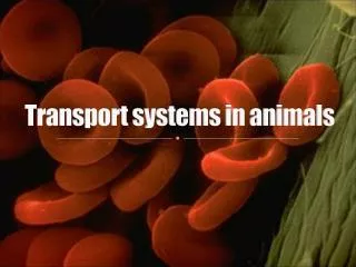
Transport in Animals By: Kadene Freckleton Teacher : Mrs. Haughton
Objectives • List and discuss briefly at least 7 main functions of the mammalian blood. • Describe the structures and functions of the following in the blood: plasma, erythrocytes, platelets and leucocytes : lymphocytes, monocytes, phagocytes (granulocytes), eosinophils, basophils, neutrophils. • Describe the structure and function of haemoglobin (briefly). • Explain the role of haemoglobin in O2 and CO2 transport.
Functions of the mammalian blood • Temperature regulation – the blood distributes heat from the deeply seated organs. This helps to maintain a constant body temperature. • Transport of gases - transports oxygen from the lungs to all parts of the body, and transports carbon dioxide produced by the cells and tissues in the reverse direction. • Transport of hormones – from the glands where there are produced to the target organs. This allows communication within the body. Eg (insulin from the pancreas to the liver) • Transport of soluble organic compounds (digested food) – from the small intestine to various parts of the body where they are stored and assimilated, and transport from storage areas to places where they are used. Eg (transport of glucose from the liver to the muscles when glycogen is converted to glucose)
Cont’d • Transport of soluble excretory materials – to organs of excretion. Eg (urea is made in the liver and transported to the kidneys for excretion). • Maintenance of a constant blood solute potential and pH as a result of plasma protein activity. • Defence against disease – blood clotting, phagocytosis, immunity.
Components of the Blood There are many substances that can be found in the blood. Some of these are proteins ( enzymes, hormones, blood clotting proteins etc.), salts (Na ions), waste products (gases, inorganic substances) and digested soluble substances. The structure of some of the other substances in the blood allows them to perform their function. These include: • Plasma • Erythrocytes • Platelets • Leucocytes- lymphocytes, monocytes, phagocytes, eosinophils, basophils, neutrophils.
Plasma The plasma is the fluid part of the blood, it is a pale straw- coloured liquid. It makes up about 55% by volume of the blood and the cells make up the other 45%. Plasma is 90% water and 10% of a variety of substances in solution and suspension. The water of the plasma is a major constituent of lymph. It transports many dissolved materials round the body and provides all the cells in the body with water. Regulation of water contents helps to regulate blood temperature and blood pressure.
Erythrocyte Erythrocytes (also known as red blood cells) are small cells that lack a true nuclei – this means they cannot reproduce. They are made in the bone marrow. They appear as biconcave discs, and are very thin. Their shape makes them very flexible and this allows them to squeeze through capillaries. They have a large surface area to volume ratio, hence efficient gaseous exchange occur. There are approximately 5 000 000 red blood cell per mm cube of blood (one drop of blood has a volume of about 50 mm cube) . Also, their life span is very short – 3 months! This is due to the lack of a nucleus to control repair processes.Red- blood cells are packed with haemoglobin. Their lack of nuclei makes more room for the haemoglobin.
Platelet Platelets are irregular shaped membrane bound cell fragments which lack nuclei. They are about one quarter the size of an erythrocyte, and are formed by special bone marrow cells. There are about 250 000 per cm cube of blood. They survive for about 5 – 9 days. Their function is to start the process of blood clotting.
Leucocytes Leucocytes (white blood cells) are present in much smaller numbers (about 7000 per mm cube of blood). They all have nuclei and play a very important role in the body’s defence mechanisms against disease. Their life span in the bloodstream is normally a few days. Leucocytes can squeeze through capillary walls to reach tissues where there are infections. They do this by a type of movement known as amoeboid movement. There are two main groups of leucocytes. They are: • Granulocyte – their cytoplasm contains granules. • Agranulocyte –their cytoplasm does not contain granules.
Granulocyte (Phagocyte) They are also called polymorphonuclear leucocytes, and make up 72% of all the white blood cells. They are made in the bone marrow by different cells that make red blood cells. Granulocytes are a type of phagocytes- they engulf invading microorganisms (phagocytosis). Granulocytes have a short life span in the circulation, typically about 6- 8 hours! There are 3 types of granulocytes: • Neutrophils • Eosinophils • Basophils
Neutrophil They make up 70% of the total number of white blood cells. They maintain a strong defensive purpose in the blood and swarm to attack invading pathogens. They can squeeze between the cells in the capillary walls, and through the intercellular spaces. Form here they move to infected areas in the body. Neutrophils have about 4 or 5 lobes. They are actively phagocytic and engulf and digest disease causing bacteria. Neutrophils that die in the line of duty release toxic chemicals to continue their protected actions. Numerous health conditions can lower the level of neutrophils levels hence raising the risk for an infection.
Eosinophil They contain cytoplasmic granules that stain red when the dye eosine is applied to them. They make up 1.5% of the total no. of white blood cells, and circulate about 8 hrs. in the blood and then migrate into the tissues. Their no. can be increased in people with allergic conditions such as asthma and hayfever. The number of eosine present in the bloodstream is under the control of hormones produced by the adrenal cortex in response to various kinds of stress.
Basophil Contains cytoplasmic granules that stain blue with basic dyes like methylene blue. They make up 0.5% of the population of the white blood cells. These cells produce heparine, anti-clotting protein and histamine- a chemical found in damaged tissues which is involved in inflammation. Inflammation stimulates repair of the damaged tissues . These cells are abundant in bronchial tissues during asthma attacks, or the tissue surrounding an insect bite or sting.
Agranulocyte They are also called mononuclear leucocytes and make up 28% of all the white blood cells. The lack cytoplasmic granules and their nucleus is either oval or bean shaped. There are two types: • Monocytes • Lymphocytes
Monocyte They are produced by the bone marrow and the lymph node and exist in the body for about 30 – 40 hrs. 4% of the body’s leucocytes are monocytes. They have a two phase existence in the body. During the first 24 hrs ( first phase) they function as a phagocyte that consumes pathogenic particles. After this 24 hr period the monocyte enters it second phase of life. It enters a tissue and is now called a macrophage (that may acquire a specific depending on its location) when it matures. They have the largest nucleus…! Together with the neutrophils they form a system of phagocytes throughout the body which acts as a first line of defence against infection.
Lymphocytes These cells are produced in the Thymus gland and Lymphoid tissue from the cells which originate in the bone marrow. Of all the white blood cells present in the body, 24% of these are lymphocytes. Lymphocytes are rounded and possess only a small quantity of cytoplasm. Amoeboid movement is limited. There are also found in the lymph and body tissues. There are two types: • T – cells • B – cells They are involved immune reactions (such as anti-body production, graft rejection and in killing tumour cells). Their life span can vary from a matter of days to up to ten years or more.
Haemoglobin Haemoglobin is found in all red blood cells, and is responsible for the transport of oxygen round the body. A haemoglobin molecule has four polypeptide chain : 2 alpha chains – 141 a.a, and two beta chains – 146 a.a. (quaternary structure). Each polypeptide chain possesses a globin polypepetide chain that is linked to haem group and is responsible for the characteristic red colour blood.
Oxygen combines with haemoglobin to form oxyhaemoglobin in high oxygen concentrations. When the concentration of oxygen is low, the bonds holding the oxygen to haemoglobin become unstable releases oxygen. Carbon Dioxide combines with the amine group- (NH2) at the end of each polypeptide chain of haemoglobin to form a neutral carbamino-haemoglobin compound.