Download
1 / 15
160 likes | 410 Vues
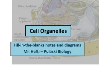
3D Interactive cell. Inner Life of the Cell (Harvard’s gorgeous animation of a cell contents doing their thing). Utah cell. The nucleus. The nuclear envelope Nuclear pores Nucleoplasm Chromatin The nucleolus. The nucleus:

E N D
3D Interactive cell Inner Life of the Cell (Harvard’s gorgeous animation of a cell contents doing their thing) Utah cell
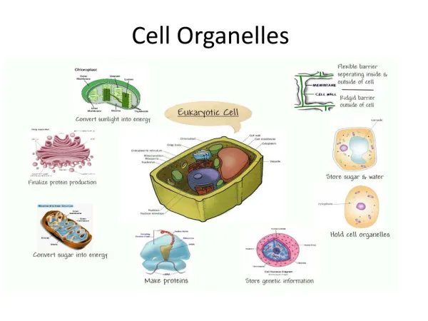
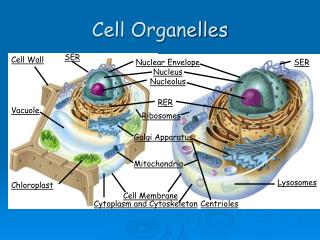
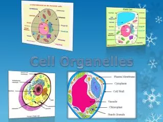
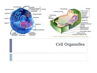
The nucleus • The nuclear envelope • Nuclear pores • Nucleoplasm • Chromatin • The nucleolus The nucleus: Acts as the control centre of the cell through the production of mRNA and protein synthesis Retains the genetic material of the cell in the form of DNA / chromosomes. Nucleolus..Manufacture rRNA and ribosomes Start the process of cell division.
Chloroplasts Role in photosynthesis. • The chloroplast envelope • The stroma • The grana • Starch grains
The mitochondrion • Double membrane • Cristae (stalked elementary particles) • The matrix (Krebs cycle)
Endoplasmic reticulum • RER • SER • Cisternae
Ribosomes Found in all cells. Role in protein synthesis 80S type = eukaryotic cells 70S type = prokaryotic cells Composed of proteins and RNA 2 sub-units. One large and one small.
Golgi apparatus • Similar to SER in structure. • Flattened stacks of cisternae. • Modifies proteins from E.R. Labels them with receptors, which directs them to their correct destination. Modified protein transported in vesicles Surface view
Lysosomes • Formed when vesicles produced from the golgi apparatus include within them enzymes (e.g. Proteases, lipases) • Up to 1.0 μm in diameter • Isolate potentially harmful enzymes from rest of cell.
Cilia • Threads which extend from cell surface. • 3-4 μm long, could be up to 10 μm. they are 0.2 μm in diameter. • Basal body embedded in cytoplasm • Basal body contains 9 sets of 3 microtubules.
Centrioles • Found in almost all animal cells. • NOT in plant cells • 0.5 μm in length and 0.2 μm in diameter. • Internal structure = 9 sets of 3 microtubules • 2 in each cell. Lie at right angles to one another near the nucleus
Cellulose Cell Wall Made of cellulose ( a polysaccharide) Several layers. Permeable Flexible Provides strength and support Pores filled with cytoplasm between adjacent cells called plasmadesmata allow movement of materials between cells
Comparing plant and animal cells Animal cells • No cell wall • No chloroplasts • Vacuoles if present – small and scattered. • Cytoplasm present throughout the cell • Nucleus anywhere in cell, but often central. • Centrioles present • Cilia or undulipodia often present • Glycogen granules used for storage Plant cells • Cell wall with pits and plasmodesmata. • Chloroplasts present • Large single vacuole. • Cytoplasm – thin layer confined to edge of cell. • Nucleus at edge of cell • No centrioles in higher plants • No cilia or undulipodia in higher plants. • Starch grains used for storage
Plenary activity: Complete short answer questions on cell organelles. Complete Q3 from Q booklet Homework: Q1 part (a) only & Q4 all parts. Due: Next lesson.