Download
1 / 80
950 likes | 1.37k Vues
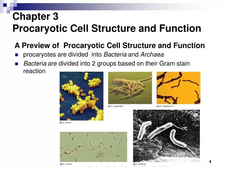
A Preview of Procaryotic Cell Structure and Function. Chapter 3 Procaryotic Cell Structure and Function. procaryotes are divided into Bacteria and Archaea Bacteria are divided into 2 groups based on their Gram stain reaction. Size, Shape, and Arrangement. cocci (s., coccus) – spheres

E N D
A Preview of Procaryotic Cell Structure and Function Chapter 3 Procaryotic Cell Structure and Function • procaryotes are divided into Bacteria and Archaea • Bacteria are divided into 2 groups based on their Gram stain reaction
Size, Shape, and Arrangement • cocci (s., coccus) – spheres • diplococci (s., diplococcus) – pairs • streptococci – chains • staphylococci – grape-like clusters • tetrads – 4 cocci in a square • sarcinae – cubic configuration of 8 cocci • bacilli (s., bacillus) – rods • coccobacilli – very short rods • vibrios – resemble rods, comma shaped • spirilla (s., spirillum) – rigid helices • spirochetes – flexible helices • mycelium – network of long, multinucleate filaments • pleomorphic – organisms that are variable in shape
largest – • 50 μm in • diameter • smallest – • 0.3 μm in • diameter
Procaryotic Cell Membranes • membranes are an absolute requirement for all living organisms • plasma membrane encompasses the cytoplasm • some procaryotes also have internal membrane systems Functions of the plasma membrane • separation of cell from its environment • selectively permeable barrier • some molecules are allowed to pass into or out of the cell • transport systems aid in movement of molecules • location of crucial metabolic processes • detection of and response to chemicals in surroundings with the aid of special receptor molecules in the membrane
Fluid Mosaic Model of Membrane Structure • Lipid bilayer in which proteins float Membrane proteins • peripheral proteins • loosely associated with the membrane and easily removed • integral proteins • embedded within the membrane and not easily removed
The asymmetry of most membrane lipids • polar ends • interact with water • hydrophilic • nonpolar ends • insoluble in water • hydrophobic Bacterial Membranes • differ from eucaryotes in lacking sterols • do contain hopanoids, sterol-like molecules • a highly organized, asymmetric system which is also flexible and dynamic
Internal Membranous Structures • plasma membrane infoldings • observed in many photosynthetic bacteria and in procaryotes with high respiratory activity • mesosomes • function not agreed upon • may be artifacts of chemical fixation of bacteria for electron microscopy
Archaeal membranes • composed of unique lipids • some have a monolayer structure instead of a bilayer structure
Cytoplasmic Matrix • substance in which nucleoid, ribosomes and inclusion bodies are suspended • lacks organelles bound by unit membranes • composed largely of water • is a major part of the protoplasm (the plasma membrane and everything within) The Procaryotic Cytoskeleton • homologs of all 3 eucaryotic cytoskeletal elements have recently been identified in Bacteria and one has been found in Archaea • functions include roles in cell division, protein localization and determination of cell shape
Inclusion bodies • granules of organic or inorganic material that are stockpiled by the cell for future use • some are enclosed by a single-layered membrane • membranes vary in composition • some made of proteins; others contain lipids Organic inclusion bodies • glycogen • polymer of glucose units • poly-β-hydroxybutyrate (PHB) • polymers of β-hydroxybutyrate • cyanophycin granules • large polypeptides containing about equal quantities of arginine and aspartic acid • carboxysomes • contain the enzyme ribulose-1,5,-bisphosphate carboxylase (Rubisco), enzyme used for CO2 fixation
gas vacuoles • found in cyanobacteria and some other aquatic procaryotes • provide buoyancy • aggregates of hollow cylindrical structures called gas vesicles
Inorganic inclusion bodies • polyphosphate granules • also called volutin granules and metachromatic granules • linear polymers of phosphates • sulfur granules • magnetosomes • contain iron in the form of magnetite • used to orient cells in magnetic fields
Ribosomes • complex structures consisting of protein and RNA • sites of protein synthesis • smaller than eucaryotic ribosomes • procaryotic ribosomes 70S • eucaryotic ribosomes 80S • S = Svedburg unit
The Nucleoid (類核體) Procaryotic chromosomes are located in the nucleoid, an area in the cytoplasm • irregularly shaped region • location of chromosome • usually 1/cell • not membrane-bound
The procaryotic chromosome • a closed circular, double-stranded DNA molecule • looped and coiled extensively • nucleoid proteins probably aid in folding • nucleoid proteins differ from histones Plasmids • usually small, closed circular DNA molecules • exist and replicate independently of chromosome • have relatively few genes present • genes on plasmids are not essential to host but may confer selective advantage (e.g., drug resistance) • curing is the loss of a plasmid • classification of plasmids based on mode of existence, spread, and function
The Bacteria Cell Wall • rigid structure that lies just outside the plasma membrane Functions of cell wall • provides characteristic shape to cell • protects the cell from osmotic lysis • may also contribute to pathogenicity • very few procaryotes lack cell walls
Cell walls of Bacteria • bacteria are divided into two major groups based on the response to gram-stain procedure. • gram-positive bacteria stain purple • gram-negative bacteria stain pink • staining reaction due to cell wall structure
Peptidoglycan (肽聚糖)(Murein, 胞壁質) Structure • meshlike polymer composed of identical subunits • contains N-acetyl glucosamine(N-乙酰葡萄糖胺) and N-acetylmuramic acid(N-乙酰胞壁酸)and several different amino acids • chains of linked peptidoglycansubunits are cross linked by peptides Diaminoacids present in peptidoglycan
Gram-Positive Cell Walls • composed primarily of peptidoglycan • may also contain large amounts of teichoic acids • some gram-positive bacteria have layer of proteins on surface of peptidoglycan Isolated gram+ cell wall • teichoic acids • polymers of glycerolor ribitol (核糖醇) joined byphosphate groups
Periplasmic Space of Gram + bacteria • lies between plasma membrane and cell wall and is smaller than that of Gram - bacteria • periplasm has relatively few proteins • enzymes secreted by Gram + bacteria are called exoenzymes Exoenzymes • perform many of the same functions that periplasmic enzymes do for gram-negative bacteria
Gram-Negative Cell Walls • consist of a thin layer of peptidoglycan surrounded by an outer membrane • outer membrane composed of lipids, lipoproteins, and lipopolysaccharide (LPS) • no teichoic acids
more complex than gram + walls • peptidoglycan is ~2-5% of wall weight • periplasmic space differs from that in gram + cells • may constitute 20-40% of cell volume • many enzymes present in periplasm • outer membrane lies outside the thin peptidoglycan layer • Braun’s lipoproteins connect outer membrane to peptidoglycan
Lipopolysaccharides (LPSs) • consists of three parts • lipid A • core polysaccharide • O side chain (O antigen) Importance of LPS • protection from host defenses (O antigen) • contributes to negative charge on cell surface (core polysaccharide) • helps stabilize outer membrane structure (lipid A) • can act as an exotoxin (lipid A)
Other Characteristics of the Outer Membrane • more permeable than plasma membrane due to presence of porin proteins and transporter proteins • porin proteins form channels through which small molecules (600-700 daltons) can pass
The Mechanism of Gram Staining • thought to involve shrinkage of the pores of the peptidoglycan layer of gram-positive cells • constriction prevents loss of crystal violet during decolorization step • thinner peptidoglycan layer and larger pores of gram-negative bacteria does not prevent loss of crystal violet
The Cell Wall and Osmotic Protection • osmotic lysis • can occur when cells are in hypotonic solutions • movement of water into cell causes swelling and lysis due to osmotic pressure • cell wall protects against osmotic lysis • lysozymebreaks the bond between N-acetyl glucosamine and N-acetylmuramic acid • penicillininhibits peptidoglycan synthesis • If cells are treated with either of the above they will lyse if they are in a hypotonic solution • protoplast – cell completely lacking cell wall • spheroplast – cell with some cell wall remaining
Archaeal cell walls • lack peptidoglycan • cell wall varies from species to species but usually consists of complex heteropolysaccharides • Methanogens have walls containing pseudomurein
Protein Secretion in Procaryotes Overview of Bacterial Protein Secretion • for both types of cells, the major pathway for transporting proteins across the membrane is the Sec-dependent pathway • all protein secretion systems require energy • numerous protein secretion pathways have been identified • four major pathways are: • Sec-dependent pathway • type II pathway • type I (ABC) protein secretion pathway • type III protein secretion pathway
Sec-Dependent Pathway • also called general secretion pathway • translocates proteins from cytoplasm across or into plasma membrane • secreted proteins synthesized as preproteins having amino-terminal signal peptide • signal peptide delays protein folding • chaperone proteins keep preproteins unfolded • secA translocates preprotein through the plasma membane • When preprotein emerges from plasma membrane a signal peptidase removes the signal peptide
Protein Secretion in Gram- bacteria • 5 secretion pathways have been identified in gram- bacteria • type II and V pathways transport proteins across outer membrane that were translocated across plasma membrane by Sec-dependent pathway • types I and III pathways are Sec-independent • type IV pathway usually functions independently of the Sec pathway
ABC Protein Secretion Pathway • also called Type I Protein Secretion Pathway • ubiquitous in procaryotes • transports proteins from cytoplasm across both plasma membrane and outer membrane • secreted proteins have C-terminal secretion signals • Gram+ bacteria use a modified version of this pathway to translocate proteins across the plasma membrane
Type II Protein Secretion Pathway • Present in a number of plant and animal pathogens • transports proteins from periplasmic across outer membrane • observed in some gram-negative bacteria, including some pathogens • complex systems consisting of up to 12-14 proteins • most are integral membrane proteins Type V Protein Secretion Pathway • Most recently discovered protein secretion system • Rely on Sec-dependent pathway to move proteins across plasma membrane • When proteins are in periplasmic space many can form a channel in outer membrane through which they transport themselves; hence they are called autotransporters
Type III Protein Secretion Pathway • secretes virulence factors of gram-negative bacteria from cytoplasm, across both plasma membrane and outer membrane, and into host cell • some type III secretion machinery is syringe-shaped • secreted proteins thought to move through a translocation channel Type IV Protein Secretion Pathway • Type IV pathways are unique because they secrete proteins and transfer DNA during conjugation • Type IV systems are made of many different proteins, some of which form a syringe-like structure
Capsules, Slime Layers, and S-Layers • capsules • usually composed of polysaccharides • well organized and not easily removed from cell • slime layers (黏液層) • similar to capsules except diffuse, unorganized and easily removed • S-layers • regularly structured layers of protein or glycoprotein • In bacteria the S layer is external to the cell wall • common among Archaea, where they may be the only structure outside the plasma membrane
Glycocalyx (醣外被) • network of polysaccharides extending from the surface of the cell • a capsule or slime layer composed of polysaccharides can also be referred to as a glycocalyx
Functions of capsules, slime layers, and S-layers • protection from host defenses (e.g., phagocytosis) • protection from harsh environmental conditions (e.g., desiccation) • attachment to surfaces More functions… • protection from viral infection or predation by bacteria • protection from chemicals in environment (e.g., detergents) • facilitate motility of gliding bacteria • protection against osmotic stress
Pili and Fimbriae • fimbriae (s., fimbria) • short, thin, hairlike, proteinaceous appendages • up to 1,000/cell • mediate attachment to surfaces • some (type IV fimbriae) required for twitching motility or gliding motility that occurs in some bacteria • sex pili (s., pilus) • similar to fimbriae except longer, thicker, and less numerous (1-10/cell) • required for mating
Patterns of Flagella Distribution • monotrichous – one flagellum • polar flagellum – flagellum at end of cell • amphitrichous – one flagellum at each end of cell • lophotrichous – cluster of flagella at one or both ends • peritrichous – spread over entire surface of cell • extends from cell surface to the tip • hollow, rigid cylinder • composed of the protein flagellin • some procaryotes have a sheath around filament
Flagellar Ultrastructure The Hook and Basal Body • hook • links filament to basal body • basal body • series of rings that drive flagellar motor
Flagellar Synthesis • an example of self-assembly • complex process involving many genes and gene products • new molecules of flagellin are transported through the hollow filament • growth is from tip, not base
The Mechanism of Flagellar Movement • flagellum rotates like a propeller • in general, counterclockwise rotation causes forward motion (run) • in general, clockwise rotation disrupts run causing a tumble (打滾) (twiddle, 轉動)
Chemotaxis (化學趨化作用) • movement towards a chemical attractant or away from a chemical repellent • concentrations of chemical attractants and chemical repellents detected by chemoreceptors on surfaces of cells
Chemotaxis Towards Attractant • in presence of attractant (b) tumbling frequency is reduced and runs in direction of attractant are longer Chemotaxis away from repellent • involves similar but opposite responses
The Bacterial Endospore • formed by some bacteria • dormant • resistant to numerous environmental conditions • heat • radiation • chemicals • desiccation What makes an endospore so resistant? • calcium (complexed with dipicolinic acid) • acid-soluble, DNA-binding proteins • dehydrated core • spore coat • DNA repair enzymes
Sporogenesis • Also called endospore formation or sporulation • normally commences when growth ceases because of lack of nutrients • complex multistage process
Transformation of dormant spores into active vegetative cells • activation • prepares spores for germination • often results from treatments like heating • germination • spore swelling • rupture of absorption of spore coat • loss of resistance • increased metabolic activity • outgrowth • emergence of vegetative cell