Download
1 / 13

E N D
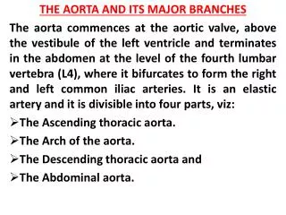
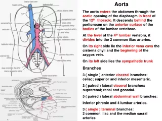
THE AORTA AND ITS MAJOR BRANCHES The aorta commences at the aortic valve, above the vestibule of the left ventricle and terminates in the abdomen at the level of the fourth lumbar vertebra (L4), where it bifurcates to form the right and left common iliac arteries. It is an elastic artery and it is divisible into four parts, viz: The Ascending thoracic aorta. The Arch of the aorta. The Descending thoracic aorta and The Abdominal aorta.
ASCENDING THORACIC AORTA: This is located in the middle mediastinum and measures 5cm long. It has the following branches through which it supplies the heart: Right coronary artery. Left coronary artery
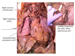
THE ARCH OF THE AORTA: This is located in the superior mediastinum. It is the continuation of the ascending aorta. Its branches are: The Brachiocephalic trunk. This gives rise to: Right Common Carotid artery. Right Subclavian artery. Left Common Carotid artery. Left Subclavian artery.
Each common carotid artery: Bifurcates to give rise to an: internal carotid artery and external carotid artery. The internal carotid continues into the cranial cavity to supply cranial contents while the external carotid supplies the head and neck region.
The Subclavian artery: The subclavian artery supplies structures in the head and neck and the thoracic regions through the following branches: Vertebral artery. The thyrocervical artery. The internal thoracic (Mammary) art. The costocervical artery The dorsal scapular artery Each Subclavian artery continues as the axillary artery at the outer margin (Lateral margin) of the first rib. The axillary artery is the main arterial supply to the upper limb.
THE DESCENDING THORACIC AORTA This artery is the direct continuation of the arch of the aorta. It passes through the posterior mediastinum and terminates posterior to the median arcuate ligament of the diaphragm at the level of the 12th thoracic vertebra (T12). At this point it continues into the abdominal cavity as the Abdominal aorta.
The branches of the Thoracic Aorta include: 9 pairs of Posterior intercostal arteries. A pair of subcostal arteries. 2 left bronchial art. 2 oesophageal art. Pericardial branches (Unknown number). Mediasternalbranches (Unknown number). A pair of Superior Phrenic art. (Right & Left).
THE ABDOMINAL AORTA This is the continuation of the descending thoracic aorta from the level of T12. It is located on the posterior abdominal wall and terminates at the level of L4. Here it bifurcates to give rise to the right and left Common iliac arteries.
The branches of the Abdominal Aorta include: Coeliactrunk. ( Foregut) Superior mesenteric art. (Midgut) Inferior mesenteric art. (Hindgut) Median sacral art. (Posterior pelvic wall) 2 Renal art. (The Kidneys) 1 Middle suprarenal art. 2 Testicular/Ovarian art. 2 Inferior phrenic art. 4 pairs of Lumbar art.
Each of the Common iliac arteries will bifurcate at the level of the sacroiliac joint to give rise toInternal and External iliac arteries. The internal iliac artery will supply the pelvic, perineal and gluteal regions. The external iliac continues as theFemoral artery beyond the inguinal ligament to supply the entire lower limb.