Download
1 / 1
10 likes | 78 Vues
A. HA detection. HA detection. GFP expression. GFP expression. B. HA detection. HA detection. GFP expression. GFP expression. Figure S1. B. A. A. C. D.

E N D
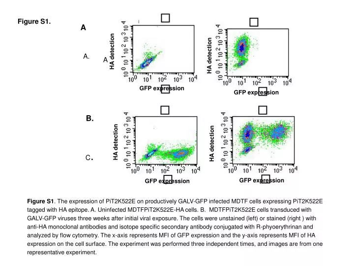
A HA detection HA detection GFP expression GFP expression B. HA detection HA detection GFP expression GFP expression Figure S1. B. A. A C. D. Figure S1. The expression of PiT2K522E on productively GALV-GFP infected MDTF cells expressing PiT2K522E tagged with HA epitope. A. Uninfected MDTFPiT2K522E-HA cells. B. MDTFPiT2K522E cells transduced with GALV-GFP viruses three weeks after initial viral exposure. The cells were unstained (left) or stained (right ) with anti-HA monoclonal antibodies and isotope specific secondary antibody conjugated with R-phyoerythrinan and analyzed by flow cytometry. The x-axis represents MFI of GFP expression and the y-axis represents MFI of HA expression on the cell surface. The experiment was performed three independent times, and images are from one representative experiment.