Download
1 / 40
400 likes | 655 Vues
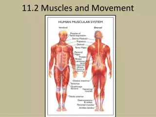
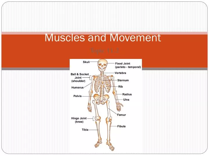
Muscles and Movement. Topic 11.2. Human Skeleton. Axial skeleton Supports the axis, or trunk of the body. Consists of : the skull, enclosing and protecting the brain the vertebral column (backbone), enclosing the spinal cord a rib cage around the lungs and heart. Human Skeleton.

E N D
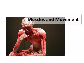
Muscles and Movement Topic 11.2
Human Skeleton • Axial skeleton • Supports the axis, or trunk of the body. • Consists of : • the skull, enclosing and protecting the brain • the vertebral column (backbone), enclosing the spinal cord • a rib cage around the lungs and heart
Human Skeleton • Appendicular skeleton • Made up of the bones of the appendages (arms and legs) and the bones that anchor the appendages to the axial skeleton. • Shoulder girdle and pelvic girdle provide a base of support for the bones of the forelimbs and hind limbs.
Human Skeleton • Distinct features: • Housing our large brain, our skull is large and flat-faced; its rounded part is the largest braincase relative to body size in the animal kingdom. • The skull is balanced atop the backbone with the spinal cord exiting directly underneath. • Our backbone is S-shaped, which helps balance the body in the vertical plane. • Pelvic girdle is short, round, and oriented vertically • Human hand is adapted for strong gripping and precise manipulation. • Our feet, with ground-touching heel, straight-facing big toe for propulsion, and shock-absorbing arches, are specialized for supporting the entire body and for bipedal walking. • Our vertical backbone bears weight unevenly, and our lower back carries much of the load. • The lower back is easily strained, especially when we bend over or lift heavy objects.
Human movement requires… • Bones • Ligaments • Muscles • Tendons • Nerves
Role of Bones • Bones • provide rigid framework against which muscles attach and against which leverage can be produced, changing the size or direction of forces generated by muscles.
Role of Ligaments • Ligaments • connect bone to bone, restricting movement at joints and helping to prevent dislocation. • Made of strong fibrous connective tissues
Role of muscles • Muscles • attach to bones via tendons, and when muscles contract, they create the forces that move bones; using leverage, small muscle contractions can produce large bone movements
Role of tendons • Tendons • attach muscles to bone.
Role of nerves • Nerves • provide a communication network along which messages can be sent signaling muscles to contract at a precise time and extent, so that movement is coordinated.
Human Skeleton • Much of the versatility of the vertebrate skeleton comes from its joints. • Strong fibrous connective tissues called ligaments hold together the bones of movable joints. • Three kinds of joints: • 1. ball-and-socket joints- HIP! • Enables us to rotate our arms and legs and move them in several planes • Protraction/retraction: forward and backwards • Abduction/adduction: sideways in and out • Rotation: circular movement • For example, where the humerus joins the shoulder girdle and also where the femur joins the pelvic girdle • 2. hinge joint- KNEE! • Permits movement in a single plane • But constrains movement from other two planes • For example, in the knee: • Flexion bends the leg • Extension straightens the leg • For example, found in the arm, elbow and also in the knee • 3. pivot joint • Enable us to rotate the forearm at the elbow. • Hinge and pivot joints in our wrists and hands enable us to make precise manipulations.
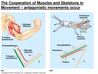
Functions of the structures of the Human Elbow • Cartilage: reduces friction between bones where they meet • Synovial fluid: lubricates joint to reduce friction • Joint capsule: seals the joint and holds in the synovial fluid • Humerus: upper arm bone: attachment of biceps and triceps • Ulna & radius: forearm bones: attachment of biceps and triceps • Biceps: attaches from humerus to ulna & radius • Triceps: attaches from humerus to ulna • Antagonism: biceps and triceps attach across elbow joint; while triceps contracts to to extend arm, biceps relaxes; conversely, while treceps relax and the biceps contract, flexing the arm
Bone • Bones are complex organs consisting of several kinds of moist, living tissues. • For example, Figure 30.4 a human humerus (upper arm bone): • Consists of a sheet of fibrous connective tissue that covers most of the outside surface.(periosteum) • Tissue helps form new bone in the event of a fracture. • A thin sheet of cartilage forms a cushion-like surface for joints, protecting the ends of bones as they move against one another. • Synovial membrane encloses the joint in synovial fluid. • Synovial fluid is formed from blood plasma and is secreted by the synovial membrane. It lubricates the joint as well as nourishing the cartilage.
Bone • For example, Figure 30.4 a human humerus (upper arm bone) continued…: • The bone itself contains living cells that secrete a surrounding material, or matrix. • Bone matrix consists of flexible fibers of the protein collagen embedded in a hard mineral made of calcium and phosphate. • The collagen keeps the bone flexible and nonbrittle, while the hard mineral matrix resists compression
Bone • For example, Figure 30.4 a human humerus (upper arm bone)continued…: • The shaft of this long bone is made of compact bone, so named because it has a dense structure. • The compact bone surrounds a central cavity with contains yellow bone marrow, which is mostly stored fat brought into the bone by the blood. • the ends, or heads, of the bone have an outer layer of compact bone and an inner layer of spongy bone, so named because it is honeycombed with small cavities. • The cavities contain red bone marrow, a specialized tissue that produces are blood cells. • Blood vessels course through channels in the bone, transporting nutrients and regulatory hormones to its cells. • Nerves paralleling the blood vessels help regulate the traffic of materials between the bone and the blood.
Skeleton and muscle interactions • Muscles interact with bones, which act as levers, to produce movement. • Muscles are connected to bones by tendons • For example, one end of the biceps muscle shown in figure 30.7 is attached by tendons to bones of the shoulder; the other end is attached across the hinge joint of the elbow—which acts as the point of support—to one of the bones in the forearm.
Skeleton and muscle interactions • The action of a muscle is always to contract, or shorten. • A muscle’s relaxation to an extended position is a passive process. • The ability to move an arm in opposite directions requires that muscles be attached to the arm bones in antagonistic pairs. • In the arm: • contraction of the biceps muscle raises the forearm. • The triceps muscle is the biceps’s antagonist. • The upper end of the triceps attaches to the shoulder, while its lower end attaches to the elbow. • The contraction of the triceps lowers the forearm, extending the biceps in the process.
Muscle tissue • Muscle tissue • Consists of bundles of long cells called muscle fibers and is the most abundant tissue in most animals. • Skeletal muscle • Attached to bones by tendons and is responsible for voluntary movements of the body. • The arrangement of the contractile units along the length of muscle cells gives them a striped or striated appearance. • Cardiac muscle • Forms the contractile tissue of the heart. • It is striated like skeletal muscle, but its cells are branched, interconnecting at specialized junctions that rapidly relay the signal to contract from cell to cell during the heartbeat. • Smooth muscle • Gets its name from its lack of striations. • Type of muscle is found in the walls of the digestive tract, urinary bladder, arteries, and other internal organs. • The cells (fibers) are shaped like spindles. They contract more slowly than skeletal muscles, but they can sustain contractions for a longer period of time.
Skeletal muscle • Skeletal muscle, which is attached to the skeleton and produces body movements, is made up of a hierarchy of smaller and smaller parallel strands. • A muscle consists of bundles of parallel muscle fibers, and each muscle fiber is a single cell with many nuclei. • Each muscle fiber is itself a bundle of smaller myofibrils. • A myofibril consists of repeating units called sarcomeres. • Skeletal muscle is called striated (striped) muscle because the sarcomeres produce alternating light and dark bands when viewed with a microscope. • Structurally, a sarcomere is the region between the two dark, narrow lines, called Z lines, in the myofibril. • Functionally, the sarcomere is the contractile apparatus in a myofibril—the muscle fiber’s fundamental unit of action.
Skeletal muscle • Sarcomere • Composed of regular arrangements of two kinds of filaments: • Thin filament • Consists of two strands of the protein actin an two strands of a regulatory protein, coiled around each other. • Light bands at the edge of the sarcomere, within light band are the Z lines that consist of proteins that connect adjuacent thin filaments • Thick filament • Contains a staggered array of multiple strands of the protein myosin. • Broad, dark band centered in the sarcomere; they are interspersed with thin filaments that project toward the center of the sarcomere. *The specific arrangement of repeating units of thin and thick filaments is directly related to the mechanics of muscle contraction.
Sliding Filament Model • Sliding-Filament model of muscle contraction: • A sarcomere contracts (shortens) when its thin filaments slide across its thick filaments. • In a contracting sarcomere: • The Z lines and the thin filaments have moved toward the middle of the sarcomere. • When the muscle is fully contracted, the thin filaments overlap in the middle of the sarcomere. • Contraction only shortens the sarcomere; it does not change the lengths of the thick and thin filaments. • A whole muscle can shorten about 35% of its resting length when all its sarcomeres contract. http://www.lab.anhb.uwa.edu.au/mb140/CorePages/Muscle/Images/Mus1ani.gif
Sliding Filament Model • What makes the thin filaments slide when a sarcomere contracts? • The key events are energy-consuming interactions between the myosin molecules of the thick filaments and the actin of the thin filaments. • Parts of the myosin, called heads, bind with specific sites on actin molecules located on the thin filaments. • In a muscle fiber at rest, these sites are covered by a regulatory protein complex of two molecules: tropomyosin and troponin • Stimulation by a motor neuron causes the binding sites to be exposed so that actin and myosis can interact. • Muscle contraction requires calcium ions (Ca2+) and energy (ATP) in order for thick and thin filaments to slide past each other.
Sliding Filament Model • Steps: • 1. The binding sites on the actin molecule (to which myosin “heads” will locate) are blocked by a complex of two molecules: tropomyosin and troponin. • 2.Prior to muscle contraction, ATP binds to the heads of the myosin molecules, priming them in an erect high energy state. • Arrival of an action potential causes a release of Ca2+ from the sarcoplasmic reticulum. • The Ca2+ binds to the troponin and causes the blocking molecules to move so that the myosin binding sites on the actin filament become exposed. • 3.The heads of the cross-bridging myosin molecules attach to the binding sites on the actin filament. • Release of energy from the hydrolysis of ATP accompanies the cross bridge formation.
Sliding Filament Model • Steps (continued…) • 4. The energy release from ATP hydrolysis causes a change in shape of the myosin cross bridge, resulting in a bending action (the power stroke). • This causes the actin filaments to slide past the myosin filaments towards the center of the sarcomere. • 5.Fresh ATP attaches to the myosin molecules, releasing them from the binding sites and repriming them for a repeat movement. They become attached further along the actin chain (closer to the Z line) as long as ATP and Ca2+ are available.
Sliding Filament Model • This sequence—detach, extend, attach, pull—occurs again and again in a contracting muscle. • Though we are only looking at one myosin head in action, a typical thick filament has about 350 heads, each of which can bind and unbind to a thin filament about five times per second. • Some myosin heads hold the thin filaments in position, while others are reaching for new binding sites. • As long as sufficient ATP is present, the process continues until the muscle is fully contracted or until the signal to contract stops.
Animation • http://www.blackwellpublishing.com/matthews/myosin.html