Download
1 / 32
330 likes | 707 Vues
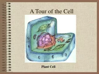
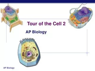
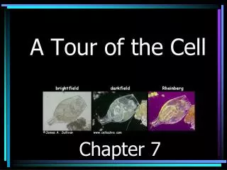
Chapter 6: A Tour of the Cell. 6.1 To study cells, biologists use microscopes and the tools of biochemistry. pp. 94 - 97. Microscopy.

E N D
6.1 To study cells, biologists use microscopes and the tools of biochemistry pp. 94 - 97
Microscopy • Light Microscope: Visible light is passed through specimen and then through glass lenses, which refract the light so that the image of the specimen is magnified. • Advantages: Does not kill specimen • Disadvantages: Cannot resolve detail finer than .2 micrometers; cellular structures are too small to be resolved by LM • Electron Microscope: Focuses beam of electrons through specimen or onto its surface. • Scanning Electron Microscope (SEM) – Electron beam scans the surface of the sample, which is usually coated with gold • Transmission Electron Microscope (TEM) – Aims electron beam through thin section of the specimen (stained with heavy metals that attach to certain organelles) to produce an image of the cell’s ultrastructure • Disadvantages: Methods used to prepare specimen often kill the cells and may introduce artifacts (illusions of structural features that don’t really exist)
Cell Fractionation • Takes apart and separates major organelles and other subcellular structures from one another. • Uses centrifuge. Spins test tubes holding mixtures of cells at various speeds. • Low speeds produce pellets with larger components. • High speeds produce pellets with smaller components. • Through fractionation, biologists discovered that mitochondria are the sites of cellular respiration.
6.2 Eukaryotic cells have internal membranes that compartmentalize their functions pp. 98 - 102
Comparing Plant and Animal Cells • In animal cells but not plant cells: • Lysosomes • Centrosomes, with centrioles • Flagella (but present in some plant sperm) • In plant cells but not animal cells: • Chloroplasts • Central vacuole • Cell wall • Plasmodesmata
6.3 The eukaryotic cell’s genetic instructions are housed in the nucleus and carried out by the ribosomes pp. 102 - 104
The Nucleus: Information Central • Nucleus contains most of the genes in the eukaryotic cell. • Some genes are found in mitochondria and chloroplasts. • Nuclear envelope encloses nucleus. Double membrane with pores. Pore complex regulates entry and exits of most proteins, macromolecules, and RNAs. • DNA -> chromatin -> chromosomes • Nucleolus: rRNA is synthesized here. Proteins are imported to the nucleolus and assembled with rRNA to make large and small ribosomal subunits.
Ribosomes: Protein Factories • Carry out protein synthesis. • Cells with high rates of protein synthesis often have large numbers of ribosomes. • Free ribosomes: suspended in the cytosol • Bound ribosomes: attached to outside of ER or nuclear envelope • Free and bound ribosomes are structurally identical and can alternate between the two roles. • Free ribosomes usually make proteins that function within cytosol. • Bound ribosomes tend to make proteins destined for • Insertion into membranes • Packaging within certain organelles • Export from the cell • Cells that specialize in protein secretion have large numbers of bound ribosomes.
6.4 The endomembrane system regulates protein traffic and performs metabolic functions in the cell pp. 104 - 108
Functions of the Endomembrane System • Synthesis of proteins • Transport of said proteins into membranes and organelles or out of the cell • Metabolism and movement of lipids • Detoxification of poisons • Includes: • Nuclear envelope • ER • Golgi • Lysosomes • Vacuoles • Plasma membrane
Endoplasmic reticulum (aka the endoplastic ridiculous . . . I AM NOT A LIPID.)
Golgi Apparatus: Shipping and Receiving Center • Products of the ER modified and stored and sent to other destinations • Cisternae • Structural polarity: cis and trans face • These are, respectively, the receiving and shipping departments • Cisternal maturation model: Products move from cis to trans and are modified in the process • Golgi manufactures certain molecules by itself - polysaccharides
Lysosomes: Digestive Compartments • Contains active hydrolytic enzymes that digest food particles. • Phagocytosis: to eat by engulfing smaller organisms. • In this way, food vacuoles fuse with lysosome, and the lysosome’s enzymes digest the food • Digestion products become nutrients for the cell • Autophagy: recycling the cell’s own organic material • Damaged organelle is surrounded by a vesicle with a double membrane. Lysosome fuses with this vesicle. Enzymes dismantle enclosed material and organic monomers are returned to the cytosol for reuse. • Tay-Sachs disease: lysosomal storage disease. Causes afflicted to lack a lipid-digesting enzyme. Brain becomes impaired by an accumulation of lipids in the cells.
Vacuoles: Diverse Maintenance Compartments • Food vacuoles: formed by phagocytosis • Contractile vacuoles: pump excess water out of the cell • Central vacuole: plants and fungi have these. Develops by coalescence of smaller vacuoles. Carries out hydrolysis but has other roles: • Reserves of organic compounds • Main repository of organic ions • Some have pigments that color the cells • Protect plant against predators by containing poisonous compounds • Plant cells enlarge as vacuole absorbs water
6.5 Mitochondria and chloroplasts change energy from one form to another pp. 109 - 111
Mitochondria: Chemical Energy Conversion • Major site of cellular respiration • Two membranes • Inner membrane folded – cristae. Two compartments: • Intermembrane space: region between inner and outer membranes • Mitochondrial matrix: enclosed by inner membrane • Contain enzymes, mitochondrial DNA, ribosomes, and ATP synthase
Chloroplasts: Capture of Light Energy • Plant organelle • Belongs to plastid family • Chlorophyll • Lens-shaped • Stacks of thylakoids. Individual stack is a granum. • Internal fluid called stroma • Double membrane divides chloroplast into three compartments: intermembrane space, stroma, and thylakoid space
Peroxisomes: Oxidation • Bounded by a single membrane • Contains enzymes that transfer hydrogen to oxygen, thereby making hydrogen peroxide • Additional enzyme converts hydrogen peroxide to water. • If peroxisome contents – that is, the hydrogen peroxide - leaked out, the cell would die • Glyoxisomes: peroxisomes found in the fat-storing tissues of plant seeds. Convert fatty acids to sugar.
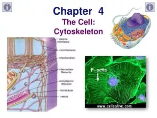
6.6 The cytoskeleton is a network of fibers that organizes structures and activities within the cell pp. 112 - 118
Roles of the Cytoskeleton • Support • Motility • Regulation • Provides anchorage for many organelles • Motility generally requires interaction between cytoskeleton and motor proteins • Cytoskeleton is composed of microtubules, microfilaments, and intermediate filaments
Microtubules • Hollow tubes; wall consists of 13 columns of tubulin molecules • 25 nm diameter with 15-nm lumen (folds) • Protein subunit is tubulin, a dimer consisting of alpha tubulin and beta tubulin. • Functions: • Maintenance of cell shape • Cell motility (as in cilia or flagella) • Chromosome movements in cell division • Organelle movements
Microfilaments • Two intertwined strands of actin • Diameter 7 nm • Protein subunit is . . . well, actin • Functions: • Maintenance of cell shape • Changes in cell shape • Muscle contraction • Cytoplasmic streaming • Cell motility (as in pseudopodia) • Cell division (cleavage furrow formation)
Intermediate Filaments • Fibrous proteins supercoiled into thicker cables • 8 – 12 nm diameter • One of several different proteins of the keratin family, depending on cell type • Functions: • Maintenance of cell shape • Anchorage of nucleus and certain other organelles • Formation of nuclear lamina
Centrosomes and Centrioles • Centrosome: Located near nucleus. Microtubule organizing center. • Centrioles: Located in the centrosome. One pair composed of nine sets of triplet microtubules arranged in a ring. • Before an animal cell divides, the centrioles replicate • Yeast cells and plant cells lack centrosomes with centrioles, but they still have microtubules.
Cilia and Flagella • Cilia are the hair-like structures on the outside of the cell. Flagella are tail-like structures. • Cilia: • Occur in large numbers on the cell surface • .25 micrometers in diameter • 2-20 micrometers long • Work more like oars – alternating power and recovery strokes perpendicular to the cilium’s axis • Flagella: • Same diameter as cilia but 10-200 micrometers long • Usually limited to just one or two per cell • Undulating motion that generates force in same direction as the flagellum’s axis.
6.7 Extracellular components and connections between cells help coordinate cellular activities pp. 118 - 122
Cell Walls of Plants • Protects plant cell • Maintains its shape • Prevents excessive uptake of water • Thicker than the plasma membrane • Composed of cellulose microfibrils, which are synthesized by cellulose synthase and secreted to the extracellular space • Perforated by plasmodesmata
The Extracellular Matrix (ECM) of Animal Cells • Main component is glycoproteins secreted by cell • Most abundant glycoprotein in animal ECM is collagen • Collagen fibers embedded in a network of proteoglycans • Some cells attached to the ECM by fibronectin • Integrins: cell surface receptor proteins. Span the membrane and bind on the cytoplasmic side to other proteins attached to microfilaments.