Download
1 / 27
270 likes | 278 Vues
Lab 1: INTRODUCTON of HISTOPTHOLOGY. 2016/2017. INTRODUCTON. Histopathology : is a branch of pathology which deals with the study of disease in a tissue section.

E N D
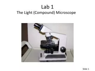
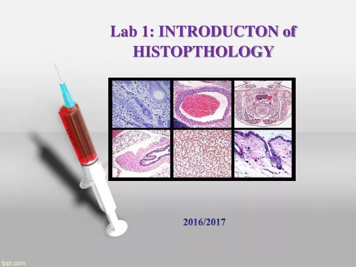
Lab 1: INTRODUCTON of HISTOPTHOLOGY 2016/2017
INTRODUCTON • Histopathology: is a branch of pathology which deals with the study of disease in a tissue section. • Histology: is the study of tissues and their structures of plants and animals, it's commonly performed by examining cells of tissues under alight microscope or electron microscope.
Pathology: is the study of diseases and of the changes that they causes changes in a person, an animal, or plant that are caused by diseases.
The term histochemistrymeans study of chemical nature of the tissue components by histological methods. The cell is the single structural unit of all tissues. The study of cell is called cytology. • A tissue is a group of cells specialized and differentiated to perform a specialized function. Collection of different type of cells forms an organ.
Types of material 1. As biopsy: A small piece of lesions or tumor which in sent for diagnosis before final removal of the lesion or the tumor Incisional biopsy. 2. If the whole of the tumor or lesion is sent for examination and diagnosis by the pathologist, it is called excisional biopsy. 3. Tissues from the autopsy are sent for the study of disease and its course, for the advancement of medicine.
Types of Histopathological Preparations 1. Whole mounts:These are preparation entire animal eg. Fungus, parasite. These preparations should be no more than 0.2-0.5 mm in thickness.
Cont. 2. Sections:The majority of the preparations in histology are sections. The tissue is cut in about 3-5 mm thick pieces processed and 5 microns thick sections are cut on a microtome. There are 2 methods of hardening the tissues. One is by freezing them and the other is by embedding them in a hard material such at paraffin wax or gelatin.
3. Smears: Smears are made from blood, bone marrow or any fluid such as pleural or ascitic fluid, blood smear and pap smear.
4. Squash: squash preparations where cells are intentionally squashed or crushed onto a slide to reveal their contents such as the study of the stages of cell division and watch chromosomes situation..
Responsibility of a technician • Specimen preservation. • Specimen labeling, logging and identification. 3. Preparation of the specimen to facilitate their gross and microscopy. 4. Record keeping.
To obtain these aims the following point need consideration. 1. As soon as the specimen is received in the laboratory, check if the specimen is properly labeled with the name, age, Hospital Registration No. and the nature of tissue to be examined and the requisition form is also duly filled. 2. Also check if the specimen is in proper fixative. Fixative should be fifteen to twenty times the volume of the specimen add fixative if not present in sufficient amount.
3. Check if the financial matters have been taken care off. 4. Make the entries in biopsy register and give the specimen a pathology number called the accession number. Note this number carefully on the requisition form as well as the container. This number will accompany the specimen everywhere.
5. If the specimen is large inform the pathologist who will make cut in the specimen so that proper fixation is done. Container should be appropriate to hold the specimen without distorting it. 6. Blocks of tissues taken for processing should be left in 10% formalin at 60°C till processing. These would be fixed in 2 hours.
7. Slides should be released for recording after consultation with the pathologist. 8. Specimens should be kept in their marked container and discarded after checking with pathologist.
9. Block must be stored at their proper number the same day. • Note the blocks have to be kept preserved for life long.
Equipment used during this course: Microtome