Download
1 / 28
320 likes | 693 Vues
Muscle Physiology. Chapter 10 . Nerve and Blood Supply. Each skeletal muscle is supplied by a nerve, artery and two veins. Each motor neuron supplies multiple muscle cells (neuromuscular junction)

E N D
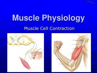
Muscle Physiology Chapter 10
Nerve and Blood Supply • Each skeletal muscle is supplied by a nerve, artery and two veins. • Each motor neuron supplies multiple muscle cells (neuromuscular junction) • Each muscle cell is supplied by one motor neuron terminal branch and is in contact with one or two capillaries. • nerve fibers & capillaries are found in the endomysium between individual cells
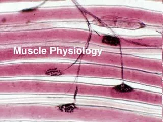
Muscle Fiber or Myofibers • Muscle cells are long, cylindrical & multinucleated • Sarcolemma = muscle cell membrane • Sarcoplasm filled with tiny threads called myofibrils & myoglobin (red-colored, oxygen-binding protein)
Transverse Tubules • T (transverse) tubules are invaginations of the sarcolemma into the center of the cell • filled with extracellular fluid • carry muscle action potentials down into cell • Mitochondria lie in rows throughout the cell • near the muscle proteins that use ATP during contraction
Myofibrils & Myofilaments • Muscle fibers are filled with threads called myofibrils separated by SR (sarcoplasmic reticulum) • Myofilaments (thick & thin filaments) are the contractile proteins of muscle
Sarcoplasmic Reticulum (SR) • System of tubular sacs similar to smooth ER in nonmuscle cells • Stores Ca+2 in a relaxed muscle • Release of Ca+2 triggers muscle contraction
Filaments and the Sarcomere • Thick and thin filaments overlap each other in a pattern that creates striations (light I bands and dark A bands) • The I band region contains only thin filaments. • They are arranged in compartments called sarcomeres, separated by Z discs. • In the overlap region, six thin filaments surround each thick filament
Thick & Thin Myofilaments • Supporting proteins (M line, titin and Z disc help anchor the thick and thin filaments in place)
The Proteins of Muscle • Myofibrils are built of 3 kinds of protein • contractile proteins • myosin and actin • regulatory proteins which turn contraction on & off • troponin and tropomyosin • structural proteins which provide proper alignment, elasticity and extensibility • titin, myomesin, nebulin and dystrophin
The Proteins of Muscle -- Myosin • Thick filaments are composed of myosin • each molecule resembles two golf clubs twisted together • myosin heads (cross bridges) extend toward the thin filaments • Held in place by the M line proteins.
The Proteins of Muscle -- Actin • Thin filaments are made of actin, troponin, & tropomyosin • The myosin-binding site on each actin molecule is covered by tropomyosin in relaxed muscle • The thin filaments are held in place by Z lines. From one Z line to the next is a sarcomere.
The Proteins of Muscle -- Titin • Titan anchors thick filament to the M line and the Z disc. • The portion of the molecule between the Z disc and the end of the thick filament can stretch to 4 times its resting length and spring back unharmed. • Role in recovery of the muscle from being stretched.
Other Structural Proteins • The M line (myomesin) connects to titin and adjacent thick filaments. • Nebulin, an inelastic protein helps align the thin filaments. • Dystrophin links thin filaments to sarcolemma and transmits the tension generated to the tendon.
Sliding Filament Mechanism Of Contraction • Myosin cross bridgespull on thin filaments • Thin filaments slide inward • Z Discs come toward each other • Sarcomeres shorten.The muscle fiber shortens. The muscle shortens • Notice :Thick & thin filaments do not change in length
How Does Contraction Begin? • Nerve impulse reaches an axon terminal & synaptic vesicles release acetylcholine (ACh) • ACh diffuses to receptors on the sarcolemma & Na+ channels open and Na+ rushes into the cell • A muscle action potential spreads over sarcolemma and down into the transverse tubules • SR releases Ca+2 into the sarcoplasm • Ca+2 binds to troponin & causes troponin-tropomyosin complex to move & reveal myosin binding sites on actin--the contraction cycle begins
Excitation - Contraction Coupling • All the steps that occur from the muscle action potential reaching the T tubule to contraction of the muscle fiber.
Contraction Cycle • Repeating sequence of events that cause the thick & thin filaments to move past each other. • 4 steps to contraction cycle • ATP hydrolysis • attachment of myosin to actin to form crossbridges • power stroke • detachment of myosin from actin • Cycle keeps repeating as long as there is ATP available & high Ca+2 level near thin filament
ATP and Myosin • Myosin heads are activated by ATP • Activated heads attach to actin & pull (power stroke) • ADP is released. (ATP released P & ADP & energy) • Thin filaments slide past the thick filaments • ATP binds to myosin head & detaches it from actin • All of these steps repeat over and over • if ATP is available & • Ca+ level near the troponin-tropomyosin complex is high
Overview: From Start to Finish • Nerve ending • Neurotransmittor • Muscle membrane • Stored Ca+2 • ATP • Muscle proteins
Relaxation • Acetylcholinesterase (AChE) breaks down ACh within the synaptic cleft • Muscle action potential ceases • Ca+2 release channels close • Active transport pumps Ca2+ back into storage in the sarcoplasmic reticulum • Calcium-binding protein (calsequestrin) helps hold Ca+2 in SR (Ca+2 concentration 10,000 times higher than in cytosol) • Tropomyosin-troponin complex recovers binding site on the actin
Rigor Mortis- Code 5 Patients (“Obviously Dead”)-Policy 4.2 • Rigor mortis is a state of muscular rigidity that begins 3-4 hours after death and lasts about 24 hours • After death, Ca+2 ions leak out of the SR and allow myosin heads to bind to actin • Since ATP synthesis has ceased, crossbridges cannot detach from actin until proteolytic enzymes begin to digest the decomposing cells.
Length of Muscle Fibers • Optimal overlap of thick & thin filaments • produces greatest number of crossbridges and the greatest amount of tension • As stretch muscle (past optimal length) • fewer cross bridges exist & less force is produced • If muscle is overly shortened (less than optimal) • fewer cross bridges exist & less force is produced • thick filaments crumpled by Z discs • Normally • resting muscle length remains between 70 to 130% of the optimum
Neuromuscular Junction (NMJ) or Synapse • NMJ = myoneural junction • end of axon nears the surface of a muscle fiber at its motor end plate region (remain separated by synaptic cleft or gap)
Events Occurring After a Nerve Signal • Arrival of nerve impulse at nerve terminal causes release of ACh from synaptic vesicles • ACh binds to receptors on muscle motor end plate opening the gated ion channels so that Na+ can rush into the muscle cell • Inside of muscle cell becomes more positive, triggering a muscle action potential that travels over the cell and down the T tubules • The release of Ca+2 from the SR is triggered and the muscle cell will shorten & generate force • Acetylcholinesterase breaks down the ACh attached to the receptors on the motor end plate so the muscle action potential will cease and the muscle cell will relax.
The Motor Unit • Motor unit = one somatic motor neuron & all the skeletal muscle cells (fibers) it stimulates • muscle fibers normally scattered throughout belly of muscle • One nerve cell supplies on average 150 muscle cells that all contract in unison. • Total strength of a contraction depends on how many motor units are activated & how large the motor units are
Motor Unit Recruitment • Motor units in a whole muscle fire asynchronously • some fibers are active others are relaxed • delays muscle fatigue so contraction can be sustained • Produces smooth muscular contraction • not series of jerky movements • Precise movements require smaller contractions • motor units must be smaller (less fibers/nerve) • Large motor units are active when large tension is needed
Home work • Watch Wiley Plus