Download
1 / 40
490 likes | 653 Vues
Histology of Blood tissue. Dr. Nabil Khouri. Learning Objectives. Be able to recognize all of the formed elements found in peripheral blood by light. Know the approximate abundance and life span of the formed elements.

E N D
Histology of Blood tissue Dr. Nabil Khouri
Learning Objectives • Be able to recognize all of the formed elements found in peripheral blood by light. • Know the approximate abundance and life span of the formed elements. • Understand the general functions of of the formed elements and major plasma proteins. • Be familiar with the general process of hematopoeisis. • Describe the organization of the bone marrow.
Major Plasma Proteins ProteinFunction Albumin Maintain colloid osmotic pressure; transport insoluble metabolites Globulins and Transport metal ions, protein- bound lipids, lipid-soluble vitamins Antibodies for host defense Complement proteins Destruction of microorganisms Clotting factors Formation of blood clots Plasma lipoproteins Transport of triglycerides and cholesterol to/from liver
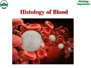
Cells of the blood • Erythrocytes(red blood cells, RBC) • Platelets (thrombocytes) • Leukocytes (white blood cells, WBC) • Granulocytes (with specific granules) • Neutrophil (~60% of WBC) • Eosinophil (~4% of WBC) • Basophil (<1% of WBC) • Agranulocytes (without specific granules) • Lymphocyte (B-cell, T-cell) (~27% of WBC) • Monocyte (~8% of WBC)
Blood smear procedure • Remember that the cells you see in a blood smear have not been sectioned. Instead you are seeing whole cells dried down on the glass • After the smear is made, it is air-dried and then stained. Common stains are Wright's stain and Giemsa stain. • The stains generally include two or more dyes, one of them a basic dye (often methylene blue) and another an acidic dye (usually eosin). • Reddish-blue azures are formed when methylene blue is oxidized. • Cells usually stain pink/red with acidic dye and nuclei stain purple/black with basic dye, while specific granules stain characteristically. • . Original Source: Junqueira's histology text, 6th ed., page 231. BloodSmear-23J91(2).tif.
Erythrocyte (red blood cell, RBC) • Life span in blood: About 120 days. • Size and shape: • biconcave disk, 8 µm diameter, 2m at thickest point, 1 m at thinnest • The shape is maintained by a cytoskeletal complex inside the plasma membrane (involving spectrin, actin and other components) • flexible: RBC’s normally bend to pass through small capillaries • LM appearance in smear: Pink circle with light center (center is thinner because of the biconcave shape). NO NUCLEUS. • TEM appearance: Solid dark gray cytoplasm, because of highly concentrated hemoglobin. • Function: Transport of oxygen and carbon dioxide • bound to hemoglobin (oxyhemoglobin and carboxyhemoglobin)
Red blood cells in a blood smear RBC Platelet
RBC, transmission electron microscopy RBC Platelet
RBCs, scanning electron microscopy Junqueira's Basic Histology, 10th edition, page 235
RBC Cytoskeleton and Membrane-Associated Proteins • Hereditary spherocytosis: defective spectrin; RBCs are fragile and destroyed in spleen leading to anemia. • A,B,O blood antigens: antigenic carbohydrate chains on extracellular domain of glycophorins • Rh antigen: multipass integral membrane protein (similar to band 3), also comprises a blood group
Platelets (thrombocytes) • Life Span: about 10 days • Shape, size, and origin: Small, biconvex disks, 2-3 µm in diameter. Non-nucleated cell fragments derived from cytoplasm of a very large cell, the megakaryocyte, in bone marrow. Platelets have a life span of about 10 days. • LM appearance in smears: Small basophilic fragments, often appearing in clusters. • Function: Platelets initiate blood clots (stop bleeding). • Not uncommonly, trauma victims, whose platelets are lost due to bleeding and related processes, require platelet transfusions. Also patients battling leukemia and other cancers – especially if they undergo transplantation with bone marrow (or related stem cell sources) – will require transfusions to make up for their platelets that are destroyed by chemotherapy and/or radiation.
Platelets Electron Microscopy Platelets Light Microscopy
Transmission electron micrographs of a platelet seen in cross section (above) and in a section in the plane of the disk (below) Fawcett's Histology, 11th edition, page 118. granule membrane tubule
Structure of a platelet • Peripheral microtubule bundle (maintains shape) • Actin and myosin (clot contraction) • Organelles facilitate clotting: • Mitochondria for ATP production • Granules contain clotting factors • Dense tubular system sequesters Ca++ for signaling (similar to SR in skeletal muscle) • Open canalicular system facilitates signaling and secretion
thromboplastin thrombin Prothrombin Thrombin Fibrinogen Fibrin Fibrin polymerization Ca++ Platelets and blood clot formation When a blood vessel wall is damaged, factors from the damaged endothelial cells and the ECM induce the clotting cascade. Platelets aggregate and release proteins for clot formation and resolution: 1. Vasoconstriction –via release of serotonin 2. Further platelet aggregation –mediated via thromboxane A2 and ADP 3. Fibrin polymerization –initiated by thromboplastin and free Ca++ 4. Clot contraction –via actin, myosin, and ATP released into the matrix of the clot 5. Clot resolution –platelet plasminogen activator (pPA, converts plasminogen into active fibrinolytic plasmin) 6. Tissue repair –platelet derived growth factor (PDGF, stimulates smooth muscle and fibroblast proliferation)
Leukocytes (white blood cells, WBC) • Granulocytes(with specific granules) • Neutrophil (~60% of WBC) • Eosinophil (~4% of WBC) • Basophil (<1% of WBC) • Agranulocytes (without specific granules) • Lymphocyte (B-cell, T-cell) (~27% of WBC) • Monocyte (~8% of WBC)
Neutrophil (polymorphonuclear leukocyte) • Life Span: < 1 week • Granulocyte with specific and non-specific granules • LM appearance in smear: About 9-12 µm in diameter (thus larger than RBC). Nucleus long and multi-lobed (usually 2-4 lobes). The Cytoplasm has small, neutrally stained specific granules. Non-specific granules are azurophilic. • Function: Primarily antibacterial • Neutrophils leave the blood and follow chemotaxic signals to sites of wounding or other inflammation, and phagocytose foreign agents such as bacteria. Pus is composed largely of dead neutrophils. • Specific granules • Type IV collagenase (aids migration) • Lactoferrin (sequesters iron) • Phospholipase A2 (leukotriene synthesis) • Lysozyme (digests bacterial cell wall) • Non-specific granules (lysosomes) • Lysozyme • Acid hydrolase • Myeloperoxidase • Elastase
Two neutrophils in a blood smear Mizoguti slide set (J). J-196. LM appearance in smear: About 9-12 µm in diameter (thus larger than RBC). Nucleus long and multi-lobed (usually 2-4 lobes). Cytoplasm has small, neutrally stained specific granules. Non-specific granules are azurophilic.
Neutrophil, transmission electron micrograph TEM appearance: Multi-lobed nucleus and numerous specific granules and lysosomes (=azurophilic granules in LM). Specific granule Lysosome (=azurophilic granule)
hydrogen peroxide hypochlorous acid Cl- superoxide O2 O2- H2O2 HOCl NADPH oxidase* superoxide dismutase myeloperoxidase Neutrophil antibacterial activity • Chemotaxis and migration (chemokine synthesis and matrix proteolysis) • Phagocytosis and bacterial destruction • Digestion via lysozymes • Production of reactive oxygen compounds (respiratory burst) • Iron sequestration via lactoferrin • Release factors to increase • inflammatory response • (and increase neutrophil production) *deficiency increases risk of persistent bacterial infections
Eosinophil • Specific granules • Major basic protein • Eosinophilic cationic protein • Neurotoxin • Histaminase • Non-specific granules (lysosomes) • Lysozyme • Acid hydrolase • Myeloperoxidase • Elastase • Life Span: < 2 weeks • Granulocyte with specific and non-specific granules • LM appearance in smear: About 10-14 µm in diameter. Bi-lobed nucleus. The cytoplasm has prominent pink/red specific granules (stained with eosin dye). If the smear is not stained properly, the granules may be brownish. • Function: • Anti-parasitic activity • Mediators of inflammatory/allergic responses in tissues • Inactivate leukotrienes and histamine secreted by basophils • Engulf and sequester antigen-antibody complexes • Inflammatory stimulus increases production/release of eosinophils from bone marrow, whereas inflammatory suppression decreases eosinophil numbers in peripheral blood.
Eosinophil in a human blood smear University of Michigan Virtual Slide Collection LM appearance in smear: About 10-14 µm in diameter. Bilobed nucleus. The cytoplasm has prominent pink/red specific granules (stained with eosin dye). If the smear is not stained properly, the granules may be brownish.
Eosinophil, transmission electron microscopy externum • TEM appearance: The specific granules are ovoid in shape, and contain a dark crystalloid body composed of major basic protein (MBP), effective against parasites. • The rest of the granule contains other anti-parasitic substances and histaminase. • The cytoplasm also contains lysosomes (=azurophilic granules). internum
Basophil • Specific granules • Histamine • Heparin • Eosinophil chemotactic factor • Phospholipids for synthesis of leukotrienes, e.g. slow-reacting substance of anaphylaxis ( SRS-A ) • Non-specific granules (lysosomes) • Lysozyme • Acid hydrolase • Myeloperoxidase • Elastase • Life Span: 1-2 years (?) • Granulocyte with specific and non-specific granules 2. LM appearance in smear: About 8-10 µm in diameter. The cytoplasm contains large, purple/black specific granules (stained with the basic dye) that are larger but not as numerous as those of eosinophils. The nucleus is usually bilobed, but usually is partially obscured by granules, which can lie over it. • Function: Allergies and anaphylaxis (hypersensitivity reaction) • Binding of antigens to membrane-bound IgE antibodies induces degranulation of specific granules, which leads to allergic reaction. • In hypersensitivity reaction, widespread vasodilation(arteriolar) and vessel leakiness induce circulatory shock. Bronchial spasms cause respiratory insufficiency; combined effect is anaphylactic shock. • Similarity to tissue mast cells: Tissue mast cells also have IgE receptors and similar (though not identical) granule content. Mast cells and basophils have a common precursor in bone marrow.
Comparison of basophil and eosinophil in a blood smear Eosinophil Basophil J.M. Velkey.
Basophil, transmission electron microscopy TEM appearance: The specific granules vary in size and shape, and have occasional myelin figures (usually formed from phospholipids). The cytoplasm also has some lysosomes (=azurophilic granules). Myelin figure Granule Erlandsen's slide set (MH). MH-2G2.
Leukocytes (white blood cells, WBC) • Granulocytes (with specific granules) • Neutrophil (~60% of WBC) • Eosinophil (~4% of WBC) • Basophil (<1% of WBC) • Agranulocytes(without specific granules) • Lymphocyte (B-cell, T-cell) (~27% of WBC) • Monocyte (~8% of WBC)
Lymphocyte • Life Span: variable (few days to several years) • LM appearance in smear:Small lymphocyte (about 90% of lymphocytes you will see) are ~8 µm in diameter, while Large lymphocytes may be up to about 15 µm. Round, dense nucleus (abundant heterochromatin). The cytoplasm of a small lymphocyte is a narrow rim around the nucleus, and when well stained is pale blue. T-lymphocytes and B-lymphocytes cannot be distinguished in a smear. • Function: Cellular and humoral immunity (more detail in the lecture and lab on lymphatic system histology). In general: • B-lymphocytes (B-cells): may differentiate into tissue plasma cells which make antibodies. Some B-cells become memory cells. • T-lymphocytes (T-cells): cytotoxic T cells and helper T cells.
Small lymphocyte in a blood smear LM appearance in smear: Small lymphocyte (about 90% of lymphocytes you will see) are ~8 µm in diameter, while large lymphocytes may be up to about 15 µm. Round, dense nucleus (abundant heterochromatin). The cytoplasm of a small lymphocyte is a narrow rim around the nucleus, and when well-stained is pale blue. Small lymphocyte
Large lymphocyte in a blood smear LM appearance in smear: Small lymphocytes (about 90% of lymphocytes you will see) are ~8 µm in diameter, while large lymphocytes may be up to about 15 µm with ovoid, dense nuclei (abundant heterochromatin). Large lymphocyte
Electron micrograph of a lymphocyte TEM appearance: The cytoplasm doesn't appear to be very active, containing mainly mitochondria and free ribosomes. Mitochondrion Centriole
Monocyte • Life Span: few days in blood, several months in connective tissue • LM appearance in smears: About 16 µm in smears, thus the largest leukocyte. Large, eccentric nucleus either oval, kidney-shaped or horseshoe-shaped, with delicate chromatin that is less dense than that of lymphocytes. Pale cytoplasm, often grayish, may contain occasional stained granules (lysosomes = azurophilic granules). Large lymphocytes may resemble monocytes, but the lymphocyte nucleus is usually more dense. • Function • Migrate into tissues and constitute mononuclear phagocyte system that help destroy foreign bodies and maintain or remodel tissues and Mediate inflammatory response Tissue macrophages Kupfer cells (liver) Osteoclasts (bone), Dust cells (lungs),Microglia (brain), • Antigen presenting cells: Dendritic Cells, Langerhans cells
Monocyte in a blood smear LM appearance in smears: About 16 µm in smears, thus the largest leukocyte. Large, eccentric nucleus either oval, kidney-shaped or horseshoe-shaped, with delicate chromatin that is less dense than that of lymphocytes. Pale cytoplasm, often grayish, may contain occasional stained granules (lysosomes = azurophilic granules). Large lymphocytes may resemble monocytes, but the lymphocyte nucleus is usually more dense.
Monocyte, transmission electron microscopy Lysosome (=azurophilic granule) Mitochondrion TEM appearance: Cytoplasm contains mitochondria and some small lysosomes. Centriole Golgi