Download
1 / 34
580 likes | 1.65k Vues
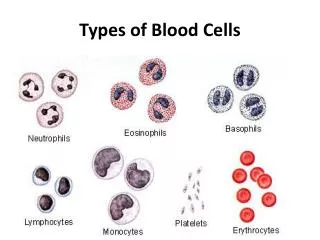
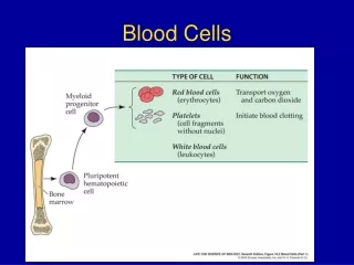
Histology of blood cells. Jeanne Adiwinata Pawitan Department of Histology FMUI. Blood – 5 lt. Specialized connective tissue – circulate – logistical support, communication Component: Blood plasma - serum Cells Erythrocytes (45%) Leucocytes – (polymorphonuclear, mononuclear) 1%

E N D
Histology of blood cells • Jeanne Adiwinata Pawitan • Department of Histology FMUI Jeanne A Pawitan
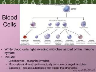
Blood – 5 lt • Specialized connective tissue – circulate – logistical support, communication • Component: • Blood plasma - serum • Cells • Erythrocytes (45%) • Leucocytes – (polymorphonuclear, mononuclear) 1% • Platelets Jeanne A Pawitan
Leukocytes • Polymorphonuclear (granular→ granulocyte) • Basophilic leukocytes (basophils) • Eosinophilic/acidophilic leukocytes (eosinophils) • Neutrophilic/heterophilic /polymorphonuclear leukocytes (neutrophils, polys) • Mononuclear (agranular→ agranulocyte) • Monocytes • Lymphocytes • B lymphocytes • T lymphocytes Jeanne A Pawitan
Leucocyte granules • Nonspecific (azurophilic) granules–0.5μm - all leucocytes • Specific granules – Giemsa/Wright • Neutrophils • Small specific granules (0.1 μm– light pink) • Tertiary granules • Eosinophils – specific granules (oblong:1-1.5 μm >< 1 μm ) - dark pink • Basophils –specific granules (0.5 μm-blue-black)-pressed to periphery →rough Jeanne A Pawitan
Azurophilic granules • = lysosomes contain: • Acid hydrolase • Myeloperoxidase • Lysozyme (antibacterial agent) • Bactericidal permeability increasing (BPI) protein • Cathepsin G • Elastase, Nonspecific collagenase → extracellular matrix →tissue damage Jeanne A Pawitan
Neutrophils • Most numerous → 60-70 % total leuco- • Ø 9-12 μm (in blood smears) • Nucleus • multilobes – chromatin threads, age ↑- lobe↑ • ♀- drumstick/Barr body - condensed, inactive, 2nd X chromosome • ↑- acute bacterial infection Jeanne A Pawitan
Neutrophil’s specific granules • Small specific granules: • Enzymes antimicrobial function • Pharmacological agent facilitate migration • Tertiary granules-neutrophil chemotactic agent→release: • Gelatinase-degrades basal lamina - facilitate migration • Cathepsins • Glycoproteins – inserted to plasmalemma – facilitate phagocytosis Jeanne A Pawitan
Neutrophil’s function • Phagocytosis – microorganism → microphage • Kill bacteria by • Enzymes • Forming reactive Oxygen compounds → die → (dead leuco, bacteria, tissue fluid) • Synthesize leucotrienes (from arachidonic acid-in their cell membrane) – aid in initiation of inflamation Jeanne A Pawitan
Phagocytosis Phagocytosis ↓ phagosome → destroyed (intracellular vacuole) ingested microorganism ↑ Azurophilic granules (lysosome) Jeanne A Pawitan
Reactive oxygen compounds • Superoxide - formed in respiratory burst (by NADPH oxidase) • Hydrogen peroxide formed from superoxide (by superoxide dismutase) • Hypochlorous acid (HOCP) formed from hydrogen peroxide and chloride ions (by myeloperoxidase) Jeanne A Pawitan
< 4 % - total leuco- Ø 10-14 μm Nucleus – bilobe Plasmalemma rec -binds to Histamin Leucotrienes Eosinophil chemotactic factor release by mast cells, baso-, neutro- → Migrate to site of Allergic reaction Parasitic worm invasion Inflammatory reaction Eosinophils Jeanne A Pawitan
Eosinophil’s specific granules • EM: • center (=internum) – crystal like –electron dense – contains: • Major basic protrein form pores in • Eosinophilic cationic protein parasites’ pellicle • Eosinophil derived neurotoxin • Externum – less electron dense- contains: • Histaminase • Peroxidase, & other enzymes Jeanne A Pawitan
Eosinophil’s function • Kill parasites-by facilitating access of superoxide, hydrogen peroxide via pores in pellicles • Release histaminase & other substances → inactivate initiators of inflamation • Histamine • Leucotriene C • Engulf Ag-Ab complex → endosomal compartment → degradation of Ag-Ab complex Jeanne A Pawitan
Basophils • < 1% total leuco- • Ø 8-10 μm • Nucleus – S shaped – masked by specific granules • Plasmalemma – surface receptors – e.g. Ig E receptors → binds IgE (from plasma cells) Jeanne A Pawitan
Basophil’s specific granules • Specific granules contain: • Heparin • Histamine • Vasodilation • Smooth muscle contraction (in the bronchial tree) • Leakiness of blood vessels • Eosinophil chemotactic factor • Neutrophil chemotactic factor • Peroxidase Jeanne A Pawitan
Basophil’s functions • IgE – Ag → • content of specific granules released • Phospholipase - phospholipids (in plasmalemma) → arachidonic acids → leukotrienes C4, D4, E4 (=slow reacting substance of anaphilaxis, SRS-A) → • similar effects with histamine, but the action is slower, and more persistent • Activate leuco- →migrate to site of Ag challenge Jeanne A Pawitan
Lymphocyte • 20-25% - total leuco- • Blood smears • Round, pleomorphic – conective tissue • Nucleus • round –slightly indented – occupies most of the cell • acentrically located • dense- heterochromatin>>> • Cytoplasm – light blue • azurophilic granules = lysosomes • Mitochondria-few, Golgi App- small, RER-few, ribosomes >>> Jeanne A Pawitan
Lymphocyte Size • Small Ø 8-10 μm • Medium Ø 12-15μm less numerous • Large Ø 15-18μm Type (functional) • B lymphocytes – 15 % - months immuno • T lymphocytes - 80 % - years cytochem • Null cells (surface markers) Jeanne A Pawitan
Lymphocyte functions • In connective tissue (not in blood) • Immune system • B lymphocyte – bone marrow immuno- • T lymphocyte – thymus (cortex) competent (maturation) →lymphoid system →mitosis →clone • Memory cells – ready for subsequent Ag chalenge • Effector cells → respond to Ag Jeanne A Pawitan
Effector cells • B cells diff→ plasma cells - Ab • T cells – cellular immune system diff → • Cytotoxic T cells (CTL, T killer cells) – contact – kill • Foreign cells • Virally altered cells • T suppressor cells →signaling molecules • T helper cells (cytokines, lymphokines) Suppression/→specific response of other cells in immnune system Jeanne A Pawitan
Monocyte • 3-8% -total leuco- • 12-15 μm • Circulation – few days → connective tissue - macrophage • Nucleus- large, acentric, kidney shaped • Chromatin network- coarse – 2 nucleoli →moth eaten/soap buble appearance • Cytoplasm – bluish gray • Azurophilic granules • Vacuole-like spaces Jeanne A Pawitan
Monocyte ME • Cytoplasm • Organels • Periphery • Microtubules • Microfilaments • Pinocytotic vesicles • Filopodia Jeanne A Pawitan
Monocyte-functions • →macrophage • Phagocytosis - phagosome – enzymatic digestion, superoxide formation→ destruction • Cells (dead, defunct) • Ag • Foreign particulate matter (bacteria) • APC – epitope (most Agnic) + class II HLA/MHC • →foreign body giant cells – large foreign particle • Cytokines • activate inflamatory response • → proliferation, maturation of other cells Jeanne A Pawitan
Platelets/thrombocyte/thromboplastids • 250,000-400,000/mm3 blood • Life span < 14 days • Disk shaped - Ø 2-4μm • Cytoplasmic fragment–megakaryocyte-bone marrow • LM • Hyalomere – periphery - clear • Granulomere - central – darker • Plasmalemma • Glycocalyx (15-20 nm) • Receptor molecules Jeanne A Pawitan
Platelets – EM - hyalomere • 10-15 microtubules (MT)-parallel → a ring – diskoid morphology • Actin, myosin monomers – associated to MT – assembly →contractile apparatus • Tubular system • Surface opening (connecting) – molecule rapid uptake & release from activated platelets • Dense tubular system – sequester Ca? >< platelet stickiness? Jeanne A Pawitan
Platelets – EM - granulomere • Few organels • Mitochondria • Peroxisomes • Glycogen deposits • Enzymes • Catabolize glycogen • Consume O2 • Generate ATP • Granules (α, δ, λ) Jeanne A Pawitan
Platelets - granules • α granules – 300-500 nm –contains • Fibrinogen • Platelet derived GF • Platelet thromboplastin • Thrombospondin • Coagulation factors • α granules content → facilitate • Vessel repair • Platelet aggregation – blood coagulation Jeanne A Pawitan
Platelets - granules • δ granules (dense bodies) – 250-300 nm - contains • Ca, ADP, ATP platelet aggregation • Serotonin, histamine & adhesion, blood • Pyrophosphatase coagulation • λ granules (lysosomes) – 200-250 nm – contains • hydrolytic enzymes →clot resorption Jeanne A Pawitan
Platelets - functions • Injury → limit hemorrhage • Platelets → activated • Tissue factors - plasma born factors – platelet derived factors → blood clot Jeanne A Pawitan
Normal Endothelium intact – inhibit platelet aggr. Prostacyclins NO2 Endothelium luminal plasmalemma – inactivate specific coagulation factor Thrombomodulin Heparin – like molecule Injury Endothelium Stop producing inhibitors Von Willebrand factor →platelet activation Tissue thromboplastin Endothelin→vasoconstr Endothelial disruption → platelet - collagen → platelet activated → → → → → → blood clot Jeanne A Pawitan
Platelet - activated • Content of granule – released • ADP platelet - sticky • thrombospondin →Platelet adhesion (to damaged vessel wall) →Platelet aggregation • Plug – block hemorrhage • Plasmalemma - platelet factor 3 = phospholipid surface – assembly of coagulation factors esp. thrombin • Arachidonic acid (plasmalemma)→ thromboxane A2 • Potent vasoconstrictor • Platelet activator Jeanne A Pawitan
Cascade of reactions in clot formation Platelet & tissue thromboplastin ↓ Prothrombin → thrombin (enzyme)→pl aggr + Ca Fibrinogen → fibrin –reticulum of clot +blood cells, platelets blot clot (thrombus) Jeanne A Pawitan
Thrombus (blot clot) formation • Erytrhrocytes – facilitate – platelet activation • Neutrophils limit platelet activation • Endothelium & thrombus size • After clot formation • 1 hour → clot contraction – ½ size → pulling edges → minimize blood lost • Repair of blood vessel → lysis of thrombus Jeanne A Pawitan
Contraction Actin monomers → thin filaments Myosin monomers → thick filaments ATP – actin-myosin filaments interact → contraction Lysis Vessel repaired → endothelium – plasminogen activators plasminogen → plasmin + λ gr. enzymes lysis of thrombus Thrombus (blood clot) Jeanne A Pawitan