Download
1 / 11
110 likes | 128 Vues
Lymphoma workshop Accessory cell and histiocytic neoplasms. Follicular dendritic cell sarcoma arising from hyaline vascular Castleman’s disease. Dr Andrew Wotherspoon Dr Larissa Sena Teixeira Mendes Histopathology Department/ Centre for Molecular Pathology Royal Marsden Hospital London, UK

E N D
Lymphoma workshopAccessory cell and histiocytic neoplasms Follicular dendritic cell sarcoma arising from hyaline vascular Castleman’s disease Dr Andrew Wotherspoon Dr Larissa Sena Teixeira Mendes Histopathology Department/ Centre for Molecular Pathology Royal Marsden Hospital London, UK Andrew.Wotherspoon@rmh.nhs.uk
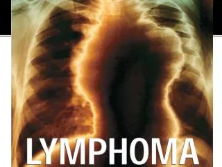
Follicular dendritic cell sarcoma Clinical history • A 26-year-old male patient presented with persistent cough in June 2013. • A chest X-ray revealed a large mediastinal mass. • CT scan showed the posterior mediastinum occupied by a large heterogeneous mass with extensive feeding vessels measuring 9,6 x 7,8cm. • There were some enlarged lymph nodes higher up the mediastinum. • PET-CT revealed an intense abnormal activity by most of the bulky soft tissue mass. The lesion appeared to be confined to the mediastinum. • The initial diagnosis was based on an EBUS-TBNA that suggested a germ cell tumour for which 2 cycles of BEP (Bleomycin+Etoposide+Cisplastin) were given, without any response. • Follicular dendritic cell sarcoma (FDCS) diagnosis was confirmed in a second biopsy of a mediastinal lymph node which also showed areas of hyaline vascular Castleman’s disease. • The patient was treated with local radiotherapy • Follow up (1y after radiotherapy treatment): patient is well and symptom free. CT scans showed post-treatment changes only, but no new measurable disease. • Background history: • Oral lichen planus since 2012 • Father was diagnosed with mantle cell lymphoma.
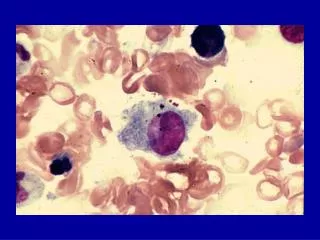
Follicular dendritic cell sarcoma Morphological findings and immunophenotypic data: • Mediastinal lymph node biopsy: • LN extensively replaced by a proliferation of pleomorphic spindle cells with a rather whorled pattern and a fibrous stroma. • The cytoplasm is pale and abundant. • Nuclei have open chromatin and multiple small nucleoli. • Some areas show a disperse proliferation of large cells with moderately atypical, ovoid vesicular and sometimes hyperchromatic nuclei • Prominent nucleoli • Scanty cytoplasm. • This sample also shows areas with regressed germinal centres, increased number of high endothelial venules with hyaline vascular changes in the interfollicular area. These features are interpreted as hyaline vascular Castleman’s disease • Bone marrow was negative for malignancies. • IHC: • Neoplastic cells were positive for CD35, CD21 and fascin. • They were negative for S100, Langerin, CD68, CD163, CD123, Cd4, CD20, Cd3, Myeloperoxidase and Lysozyme.
Follicular dendritic cell sarcoma Proposed diagnosis Follicular dendritic cell sarcoma arising from Hyaline vascular Castleman’s disease (HVCD)
Follicular dendritic cell sarcoma Interesting features: • This case illustrates the full spectrum of lesions involving histiocytes with progression from HVCD to dysplastic follicular dendritic cells to overt neoplasia (FDC sarcoma) which harbours areas showing pleomorphic features