Download
1 / 35
360 likes | 961 Vues
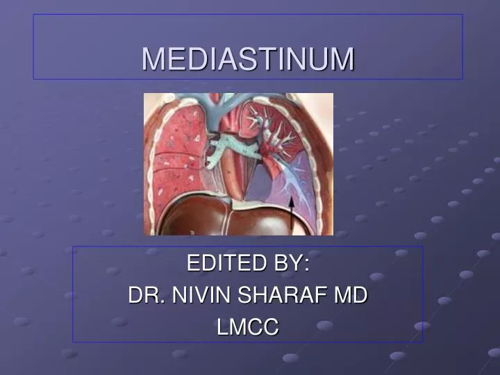
MEDIASTINUM. EDITED BY: DR. NIVIN SHARAF MD LMCC. OBJECTIVES. By the end of this lecture the students should be able to : Define mediastinum. Enlist the divisions of mediastinum. Describe the boundaries and contents of mediastinum. MEDIASTINUM. Central compartment of the thoracic cavity

E N D
MEDIASTINUM EDITED BY: DR. NIVIN SHARAF MD LMCC
OBJECTIVES By the end of this lecture the students should be able to: Define mediastinum. Enlist the divisions of mediastinum. Describe the boundaries and contents of mediastinum.
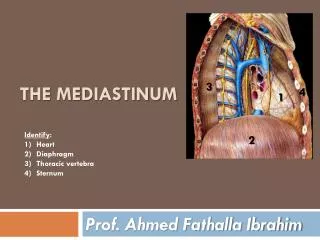
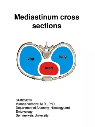
MEDIASTINUM Central compartment of the thoracic cavity Covered by mediastinal pleura Contents: all thoracic viscera EXCEPT lungs Extent: Superior - thoracic inlet Inferior - diaphragm Anterior - sternum & costal cartilages Posterior- bodies of thoracic vertebrae
MEDIASTINUM Surrounded by blood and lymphatic vessels Lymph nodes, nerves and adipose tissues Looseness of structures enable mediastinum to accommodate changes in movement, volume & pressure in the thoracic cavity
Divisions of the Mediastinum SUPERIOR MEDIASTINUM Superior - thoracic inlet Inferior - transverse thoracic plane Anterior - sternal angle Posterior - IV disc T4 & T5 INFERIOR MEDIASTINUM Superior – transverse thoracic plane Inferior - diaphragm
Divisions of the Mediastinum INFERIOR MEDIASTINUM a. ANTERIOR MEDIASTINUM - contains thymus remnant, lymph nodes & fats b. MIDDLE MEDIASTINUM - contains the heart & great vessels c. POSTERIOR MEDIASTINUM - contains esophagus, great vessels,vagus nerves & symphathetic trunks
Superior Mediastinum Contents: (Anterior – Posterior) 1. Thymus gland - primary lymphoid organ located behind manubrium - puberty undergoes gradual involution 2. Great Vessels Brachiocephalic Veins Superior Vena Cava - formed at level of 1st right costal cartilage - enters right atrium at level of 3rd right costal cartilage
Superior Mediastinum Contents: Arch of the Aorta - starts behind 2nd right SC joint - ends at 2nd left SC joint Branches: Brachiocephalic Trunk Left Common Carotid Artery Left Subclavian Artery
Aortic Arch • Passes upwards from the sternal angle behind the manubrium, backwards and to the left of the 4th Thoracic vertebra • BRANCHES SUPPLY UL, HEAD, and NECK
Ligamentum arteriosum Aortic Arch is connected inferiorly to the left pulmonary artery by the Ligamentum arteriosum”fibrous remnant of ductus arteriosus”
Superior Mediastinum Contents: 3. Nerves Vagus & Phrenic Nerves Cardiac Plexus of Nerves Left Recurrent Laryngeal Nerve 4. Trachea 5. Esophagus 6. Thoracic Duct 7. Prevertebral Muscles
Anterior Mediastinum Smallest subdivision of the Inferior Mediastinum Boundaries: Anterior : body of sternum & trans thoracis muscles Posterior : pericardium Contents: Loose CT (Sternopericardial Ligament) Adipose tissue Lymphatic Vessels & lymph nodes Branches of Internal Thoracic Vessels
Posterior Mediastinum Boundaries: Anterior - Pericardium & Diaphragm Posterior - T5 to T12 vertebrae Contents: Thoracic Aorta Esophagus & Plexus Thoracic Duct Thoracic Sympathetic Trunks Post Mediastinal LN Thoracic Splanchnic Nerve Azygos & Hemiazygos Veins
Thoracic Aorta Branches: 1. Bronchial Arteries 5. Esophageal Arteries 2. Pericardial Arteries 6. Mediastinal Arteries 3. Post. Intercostal Arteries 7. Subcostal Arteries 4. Superior Phrenic Arteries
Esophagus Course: Superior to Posterior Mediastinum Located behind: Arch of Aorta Pericardium & Left Atrium Enters Esophageal Hiatus of the Diaphragm at level of T10 Anatomic Impressions or “Constrictions”: 1. Crossing with Aortic Arch 2. Crossing with Left Main Bronchus 3. Diaphragmatic Hiatus
Thoracic Duct Largest lymphatic channel in the body Originates from Cisterna Chyle in the abdomen & passes thru aortic hiatus of diaphragm at level of T12 Relations: Posterior : bodies of inferior 7 thoracic vertebrae Anterior : Esophagus Left : Thoracic Aorta Right : Azygos Vein Conveys lymph from: Lower extremities Left side of thorax Pelvic Cavity Left side of H & N Abdominal Cavity Left upper limb
Lymph Nodes of the Posterior Mediastinum Posterior Mediastinal Lymph Nodes - receives lymph from esophagus, posterior aspect of the pericardium & diaphragm & middle posterior ICS
Azygos Venous System of the Posterior Mediastinum Drains the back & thoracoabdominal walls and the mediastinal viscera Azygos Vein - forms a collateral pathway b/w SVC & IVC - passes to the right side of inferior 8 thoracic vertebrae - arches over the root of the right lung to enter the SVC - receives posterior intercostal veins, mediastinal, esophageal & bronchial veins
Azygos Venous System of Posterior Mediastinum Hemiazygos Vein - arises on left side of the vertebral column to level of T9 - receives the inferior 3 PIV, inferior esophageal veins, small mediastinal veins Accessory Hemiazygos Vein - starts at medial end of 4th or 5th ICS - descends on left of VC from T5 thru T8 - crosses to the right to join the Azygos Vein - receives 4th thru 8th IC Veins - communicates with Superior IC Vein which drains 1st thru 3rd ICS
Nerves of Posterior Mediastinum Thoracic Sympathetic Trunks - lie against heads of ribs in superior thorax costovertebral joints in midthorax sides of vertebral bodies in lower thorax Lower Thoracic Splanchnic Nerves (Greater, Lesser and Least SN) - presynaptic fibers from 5th thru 12th sympathetic ganglia - sympathetic innervation for most abdominal viscera
References Illustrated Clinical Anatomy by:Peter Abrahams Clinical Anatomy by Systems for Richard S Snell Pages Gray’s Anatomy for students