Download

1 / 49
490 likes | 603 Vues
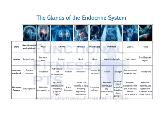
The Endocrine System: Part 2. Figure 16.9 The thyroid gland. Hyoid bone. Colloid-filled follicles. Epiglottis. Thyroid cartilage. Follicular cells. Superior thyroid artery. Common carotid artery. Inferior thyroid artery. Isthmus of thyroid gland. Trachea. Left subclavian artery.

E N D
Figure 16.9 The thyroid gland. Hyoid bone Colloid-filled follicles Epiglottis Thyroid cartilage Follicular cells Superior thyroid artery Common carotid artery Inferior thyroid artery Isthmus of thyroid gland Trachea Left subclavian artery Left lateral lobe of thyroid gland Aorta Parafollicular cells Gross anatomy of the thyroid gland, anterior view Photomicrograph of thyroid gland follicles (145x) MDufilho
Thyroid Hormone (TH) • Actually two related compounds • T4 (thyroxine); has 2 tyrosine molecules + 4 bound iodine atoms • T3 (triiodothyronine); has 2 tyrosines + 3 bound iodine atoms • Affects virtually every cell in body MDufilho
Thyroid Hormone • Major metabolic hormone • Increases metabolic rate and heat production (calorigenic effect) • Regulation of tissue growth and development • Development of skeletal and nervous systems • Reproductive capabilities • Maintenance of blood pressure MDufilho
Figure 16.10 Synthesis of thyroid hormone. Slide 1 Thyroid follicular cells Colloid 1 Thyroglobulin is synthesized and discharged into the follicle lumen. Tyrosines (part of thyroglobulin molecule) Capillary 4 Iodine is attached to tyrosine in colloid, forming DIT and MIT. Golgi apparatus Rough ER Thyro- globulin colloid Iodine DIT MIT 3 Iodide is oxidized to iodine. 2 Iodide (I−) Iodide (I–) is trapped (actively transported in). T4 5 Iodinated tyrosines are linked together to form T3 and T4. T3 Lysosome T4 6 Thyroglobulin colloid is endocytosed and combined with a lysosome. T3 7 Lysosomal enzymes cleave T4 and T3 from thyroglobulin and hormones diffuse into bloodstream. Colloid in lumen of follicle T4 T3 To peripheral tissues MDufilho
Transport and Regulation of TH • T4 and T3 transported by thyroxine-binding globulins (TBGs) • Both bind to target receptors, but T3 is ten times more active than T4 • Peripheral tissues convert T4 to T3 MDufilho
Transport and Regulation of TH • Negative feedback regulation of TH release • Rising TH levels provide negative feedback inhibition on release of TSH • Hypothalamic thyrotropin-releasing hormone (TRH) can overcome negative feedback during pregnancy or exposure to cold MDufilho
Homeostatic Imbalances of TH • Hyposecretion in adults—myxedema; goiter if due to lack of iodine • Hyposecretion in infants—cretinism • Hypersecretion—Graves' disease MDufilho
Figure 16.11 Thyroid disorders. MDufilho
Parathyroid Glands • Four to eight tiny glands embedded in posterior aspect of thyroid • Contain oxyphil cells (function unknown) and parathyroid cells that secrete parathyroid hormone (PTH) orparathormone • PTH—most important hormone in Ca2+ homeostasis MDufilho
Figure 16.12 The parathyroid glands. Pharynx (posterior aspect) Capillary Parathyroid cells (secrete parathyroid hormone) Thyroid gland Parathyroid glands Esophagus Oxyphil cells Trachea MDufilho
Figure 16.13 Effects of parathyroid hormone on bone, the kidneys, and the intestine. Hypocalcemia (low blood Ca2+) PTH release from parathyroid gland Activation of vitamin D by kidney Ca2+ reabsorption in kidney tubule Osteoclast activity in bone causes Ca2+ and PO43- release into blood Ca2+ absorption from food in small intestine Ca2+ in blood Initial stimulus Physiological response MDufilho Result
Homeostatic Imbalances of PTH • Hyperparathyroidism due to tumor • Bones soften and deform • Elevated Ca2+ depresses nervous system and contributes to formation of kidney stones • Hypoparathyroidism following gland trauma or removal or dietary magnesium deficiency • Results in tetany, respiratory paralysis, and death MDufilho
Adrenal (Suprarenal) Glands • Paired, pyramid-shaped organs atop kidneys • Structurally and functionally are two glands in one • Adrenal medulla—nervous tissue; part of sympathetic nervous system • Adrenal cortex—three layers of glandular tissue that synthesize and secrete corticosteroids MDufilho
Adrenal Cortex • Three layers and the corticosteroids produced • Zona glomerulosa—mineralocorticoids (chiefly aldosterone • Zona fasciculata—glucocorticoids (chiefly cortisol) • Zona reticularis—sex hormones, or gonadocorticoids (chiefly androgens) MDufilho
Figure 16.14 Microscopic structure of the adrenal gland. Hormones secreted Capsule Zona glomerulosa Aldosterone Zona fasciculata Adrenal gland Cortex • Medulla • Cortex Cortisol and androgens Kidney Zona reticularis Adrenal medulla Medulla Epinephrine and norepinephrine Photomicrograph (115x) Drawing of the histology of the adrenal cortex and a portion of the adrenal medulla MDufilho
Mineralocorticoids • Regulate electrolytes (primarily Na+ and K+) in ECF • Importance of Na+: affects ECF volume, blood volume, blood pressure, levels of other ions • Importance of K+: sets RMP of cells • Aldosterone most potent mineralocorticoid • Stimulates Na+ reabsorption and water retention by kidneys; elimination of K+ MDufilho
Mechanisms of Aldosterone Secretion • Renin-angiotensin-aldosterone mechanism: decreased blood pressure stimulates kidneys to release renin triggers formation of angiotensin II, a potent stimulator of aldosterone release • Plasma concentration of K+: increased K+ directly influences zona glomerulosa cells to release aldosterone • ACTH: causes small increases of aldosterone during stress • Atrial natriuretic peptide (ANP): blocks renin and aldosterone secretion to decrease blood pressure MDufilho
Figure 16.15 Major mechanisms controlling aldosterone release from the adrenal cortex. Primary regulators Other factors Stress Blood volume and/or blood pressure K+ in blood Blood pressure and/or blood volume Hypo- thalamus Heart Kidney CRH Direct stimulating effect Anterior pituitary Renin Initiates cascade that produces Atrial natriuretic peptide (ANP) ACTH Angiotensin II Inhibitory effect Zona glomerulosa of adrenal cortex Enhanced secretion of aldosterone Targets kidney tubules Absorption of Na+ and water; increased K+ excretion Blood volume and/or blood pressure MDufilho
Homeostatic Imbalances of Aldosterone • Aldosteronism—hypersecretion due to adrenal tumors • Hypertension and edema due to excessive Na+ • Excretion of K+ leading to abnormal function of neurons and muscle MDufilho
Glucocorticoids • Keep blood glucose levels relatively constant • Maintain blood pressure by increasing action of vasoconstrictors • Cortisol (hydrocortisone) • Only one in significant amounts in humans • Cortisone • Corticosterone MDufilho
Glucocorticoids: Cortisol • Released in response to ACTH, patterns of eating and activity, and stress • Prime metabolic effect is gluconeogenesis—formation of glucose from fats and proteins • Promotes rises in blood glucose, fatty acids, and amino acids • "Saves" glucose for brain • Enhances vasoconstriction rise in blood pressure to quickly distribute nutrients to cells MDufilho
Homeostatic Imbalances of Glucocorticoids • Hypersecretion—Cushing's syndrome/disease • Depresses cartilage and bone formation • Inhibits inflammation • Depresses immune system • Disrupts cardiovascular, neural, and gastrointestinal function • Hyposecretion—Addison's disease • Also involves deficits in mineralocorticoids • Decrease in glucose and Na+ levels • Weight loss, severe dehydration, and hypotension MDufilho
Figure 16.16 The effects of excess glucocorticoid. Patient before onset. Same patient with Cushing’s syndrome. The white arrow shows the characteristic “buffalo hump” of fat on the upper back. MDufilho
Gonadocorticoids (Sex Hormones) • Most weak androgens (male sex hormones) converted to testosterone in tissue cells, some to estrogens • May contribute to • Onset of puberty • Appearance of secondary sex characteristics • Sex drive in women • Estrogens in postmenopausal women MDufilho
Gonadocorticoids • Hypersecretion • Adrenogenital syndrome (masculinization) • Not noticeable in adult males • Females and prepubertal males • Boys – reproductive organs mature; secondary sex characteristics emerge early • Females – beard, masculine pattern of body hair; clitoris resembles small penis MDufilho
Adrenal Medulla • Medullary chromaffin cells synthesize epinephrine (80%) and norepinephrine (20%) • Effects • Vasoconstriction • Increased heart rate • Increased blood glucose levels • Blood diverted to brain, heart, and skeletal muscle MDufilho
Adrenal Medulla • Responses brief • Epinephrine stimulates metabolic activities, bronchial dilation, and blood flow to skeletal muscles and heart • Norepinephrine influences peripheral vasoconstriction and blood pressure MDufilho
Adrenal Medulla • Hypersecretion • Hyperglycemia, increased metabolic rate, rapid heartbeat and palpitations, hypertension, intense nervousness, sweating • Hyposecretion • Not problematic • Adrenal catecholamines not essential to life MDufilho
Figure 16.17 Stress and the adrenal gland. Short-term stress Prolonged stress Stress Nerve impulses Hypothalamus CRH (corticotropin- releasing hormone) Spinal cord Corticotropic cells of anterior pituitary Preganglionic sympathetic fibers To target in blood Adrenal cortex (secretes steroid hormones) Adrenal medulla (secretes amino acid– based hormones) ACTH Catecholamines (epinephrine and norepinephrine) Mineralocorticoids Glucocorticoids Long-term stress response Short-term stress response • Heart rate increases • Kidneys retain sodium and water • Proteins and fats converted to glucose or broken down for energy • Blood pressure increases • Bronchioles dilate • Blood volume and blood pressure rise • Liver converts glycogen to glucose and releases glucose to blood • Blood glucose increases • Immune system supressed • Blood flow changes, reducing digestive system activity and urine output • Metabolic rate increases MDufilho
Pineal Gland • Small gland hanging from roof of third ventricle • Pinealocytes secrete melatonin, derived from serotonin • Melatonin may affect • Timing of sexual maturation and puberty • Day/night cycles • Physiological processes that show rhythmic variations (body temperature, sleep, appetite) • Production of antioxidant and detoxification molecules in cells MDufilho
Pancreas • Triangular gland partially behind stomach • Has both exocrine and endocrine cells • Acinar cells (exocrine) produce enzyme-rich juice for digestion • Pancreatic islets (islets of Langerhans)contain endocrine cells • Alpha () cells produce glucagon (hyperglycemic hormone) • Beta () cells produce insulin (hypoglycemic hormone) MDufilho
Figure 16.18 Photomicrograph of differentially stained pancreatic tissue. Pancreatic islet • (Glucagon- producing) cells • (Insulin- producing) cells Pancreatic acinar cells (exocrine) MDufilho
Glucagon • Major target—liver • Causes increased blood glucose levels • Effects • Glycogenolysis—breakdown of glycogen to glucose • Gluconeogenesis—synthesis of glucose from lactic acid and noncarbohydrates • Release of glucose to blood MDufilho
Insulin • Effects of insulin • Lowers blood glucose levels • Enhances membrane transport of glucose into fat and muscle cells • Inhibits glycogenolysis and gluconeogenesis • Participates in neuronal development and learning and memory • Not needed for glucose uptake in liver, kidney or brain MDufilho
Figure 16.19 Insulin and glucagon from the pancreas regulate blood glucose levels. Stimulates glucose uptake by cells Tissue cells Insulin Stimulates glycogen formationw Pancreas Glucose Glycogen Blood glucose falls to normal range. Liver IMBALANCE Stimulus Blood glucose level BALANCE: Normal blood glucose level (about 90 mg/100 ml) Stimulus Blood glucose level IMBALANCE Blood glucose rises to normal range. Pancreas Glycogen Glucose Liver Stimulates glycogen breakdown Glucagon MDufilho
Homeostatic Imbalances of Insulin • Diabetes mellitus (DM) • Due to hyposecretion (type 1) or hypoactivity (type 2) of insulin • Three cardinal signs of DM • Polyuria—huge urine output • Glucose acts as osmotic diuretic • Polydipsia—excessive thirst • From water loss due to polyuria • Polyphagia—excessive hunger and food consumption • Cells cannot take up glucose; are "starving" MDufilho
Type 1 - IDDM • 10% of DM cases • Early onset • No insulin produced • Treatment – testing and insulin injections • Long term problems MDufilho
Type II - NIDDM • 90% of DM cases • Develops after age 40 • Causes • Produce insulin but receptors are reduced or non-responsive • Symptoms • Consequences MDufilho
Homeostatic Imbalances of Insulin • Hyperinsulinism: • Excessive insulin secretion • Causes hypoglycemia • Low blood glucose levels • Anxiety, nervousness, disorientation, unconsciousness, even death • Treated by sugar ingestion • Gestational Diabetes • Elevated levels of estrogen and progesterone make target cells resistant to insulin • Placenta releases insulinase – breakdown of insulin MDufilho
What if???? • Type I – IDDM and you eat a lot of carbs but do not take insulin • Type I – Give insulin shot but do not eat. • Type II – NIDDM – eat a big Thanksgiving meal • Tumor affecting posterior pituitary – inhibits secretion of ? MDufilho
Other Hormone-producing Structures • Adipose tissue • Leptin – appetite control; stimulates increased energy expenditure • Resistin – insulin antagonist • Adiponectin – enhances sensitivity to insulin MDufilho
Other Hormone-producing Structures • Enteroendocrine cells of gastrointestinal tract • Gastrin stimulates release of HCl • Secretin stimulates liver and pancreas • Cholecystokinin stimulates pancreas, gallbladder, and hepatopancreatic sphincter • Serotonin acts as paracrine MDufilho
Other Hormone-producing Structures • Heart • Atrial natriuretic peptide (ANP) decreases blood Na+ concentration, therefore blood pressure and blood volume • Kidneys • Erythropoietin signals production of red blood cells • Renin initiates the renin-angiotensin-aldosterone mechanism MDufilho
Other Hormone-producing Structures • Skeleton (osteoblasts) • Osteocalcin • Prods pancreas to secrete more insulin; restricts fat storage; improves glucose handling; reduces body fat • Activated by insulin • Low levels of osteocalcin in type 2 diabetes – perhaps increasing levels may be new treatment • Skin • Cholecalciferol, precursor of vitamin D MDufilho
Other Hormone-producing Structures • Thymus • Large in infants and children; shrinks as age • Thymulin, thymopoietins, and thymosins • May be involved in normal development of T lymphocytes in immune response • Classified as hormones; act as paracrines MDufilho
Developmental Aspects • Hormone-producing glands arise from all three germ layers • Most endocrine organs operate well until old age • Exposure to pesticides, industrial chemicals, arsenic, dioxin, and soil and water pollutants disrupts hormone function • Sex hormones, thyroid hormone, and glucocorticoids are vulnerable to the effects of pollutants • Interference with glucocorticoids may help explain high cancer rates in certain areas MDufilho
Developmental Aspects • Ovaries undergo significant changes with age and become unresponsive to gonadotropins; problems associated with estrogen deficiency occur • Testosterone also diminishes with age, but effect is not usually seen until very old age MDufilho
Developmental Aspects • GH levels decline with age - accounts for muscle atrophy with age • TH declines with age, contributing to lower basal metabolic rates • PTH levels remain fairly constant with age, but lack of estrogen in older women makes them more vulnerable to bone-demineralizing effects of PTH MDufilho