Download
1 / 96
960 likes | 1.11k Vues
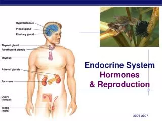
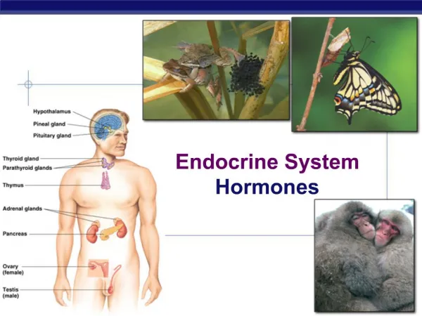
Endocrine System Part 2, Hormones. http://www.elmhurst.edu. Much of the text material is from, “Principles of Anatomy and Physiology, 12th edition” by Gerald J. Tortora and Bryan Derrickson (2009). I don’t claim authorship. Other sources are noted when they are used.

E N D
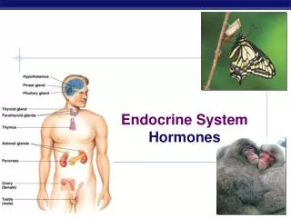
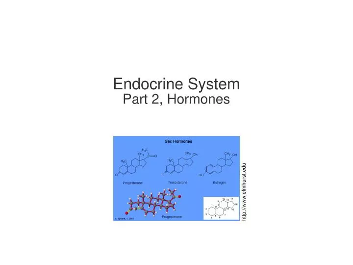
Endocrine SystemPart 2, Hormones http://www.elmhurst.edu
Much of the text material is from, “Principles of Anatomy and Physiology, 12th edition” by Gerald J. Tortora and Bryan Derrickson (2009). I don’t claim authorship. Other sources are noted when they are used. Mapping of the lecture slides to the 13th edition is provided in the supplement.
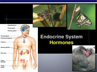
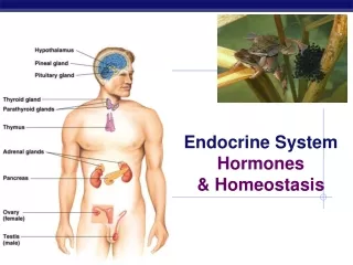
Outline • Hypothalamus and pituitary gland • Anterior pituitary • Posterior pituitary • Endocrine glands and their actions • Other endocrine organs and tissues • Stress response • Aging
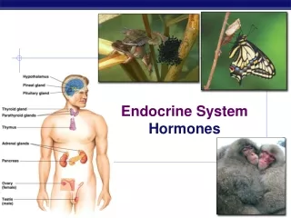
Hypothalamus and Pituitary Gland • The pituitary gland is also known as the master gland because it releases hormones that control other endocrine glands of the body. • It is regulated by the hypothalamus, the major link between the ner-vous and endocrine systems. • Cells in the hypothalamus and pituitary gland synthesize and secrete hormones. • The hormones are involved in growth and development, metabolism, homeostatis, and sexual reproduction. Chapter 18, page 650
Pituitary Structure • The pituitary gland is a pea-shaped structure located inferior to the hypothalamus, and measures about 13mm in diameter. • It is attached to the hypothalamus by the infundibulum, or pituitary stalk. • The pituitary has two major regions: 1) anterior lobe or adenohypo-physis, and 2) posterior lobe or neurohyphophysis. • The anterior lobe is about three-quarters of the total mass of the pitui-tary gland. Figure 18.5 Chapter 18, page 650
Location Magnetic resonance image (MRI) http://3d-brain.ki.se/mri
Anterior Pituitary • The anterior lobe of the pituitary gland secretes hormones that regu-late many body functions. • The secretion of hormones from the anterior pituitary is controlled by releasing hormones and inhibiting hormones from the hypothalamus. Figure 18.5 Chapter 18, page 650
Hypophyseal Portal System • Releasing hormones and inhibiting hormones reach the anterior pitui-tary through the hypophyseal portal system. • Blood in this system flows: • Through a capillary network in the hypothalamus, • Into a portal vein that transits the pituitary stalk, and • Then through a second capillary network in the anterior pituitary. • This arrangement enables chemical control of the anterior pituitary by the hypothalamus. Figure 18.5 Chapter 18, page 650
Anterior Pituitary Control Hypothalamus Hypophyseal portal system Releasing hormones and inhibiting hormones Anterior pituitary General blood circulation Hormones that affect target tissues in the body Target tissues
Hormone Entry into Blood Circulation • In response to releasing hormones, hormones secreted by cells of the anterior pituitary are secreted into the anterior hypophyseal veins. • Pituitary hormones enter general blood circulation to exert their effects on target tissues of the body. Figure 18.5 Chapter 18, page 652
Anterior Pituitary Hormones Anterior pituitary General blood circulation Human growth hormone—hGH Thyroid-stimulating hormone—TSH Follicle-stimulating hormone—FSH Luteinizing hormone—LH Prolactin—PRL Adrenocorticotropic hormone—ACTH
Neurosecretory Cells • The anterior pituitary has specific groups of cells that produce the six hormones (each group ends in -troph). • Somatatrophs synthesize and secrete human growth hormone (hGH) to stimulate certain body tissues to release insulin-like growth factors (IGFs). • The growth factors promote body growth and regulate certain aspects of metabolism. • Thyrotrophs synthesize and secrete thyroid-stimulating hormone (TSH) to control hormonal secretions and other activities of the thyroid gland. Figure 18.5 Table 18.3 Chapter 18, page 652
Neurosecretory Cells (continued) • Gonadotrophs synthesize and secrete follicle-stimulating hormone (FSH) and luteinizing hormone (LH) that act on the female and male gonads. • The hormones stimulate the secretion of estrogens and proges-terone, and promote the maturation of the oocytes (eggs) in the ovaries. • They stimulate testosterone secretion and sperm production in the testes. • Lactotrophs synthesize and secrete prolactin (PRL) to stimulate milk production in the mammary glands. Table 18.3 Chapter 18, page 652
Neurosecretory Cells (continued) • Corticotrophs secrete adrenocorticotropic hormone (ACTH), also known as corticotropin. • ACTH stimulates the adrenal cortex—the outer tissue layers of the adre-nal gland—to secrete glucocorticoids including cortisol. Table 18.3 Chapter 18, page 652
Control of Hormonal Secretions • Two mechanisms control the secretion of hormones from the anterior pituitary: • Neurosecretory cells in the hypothalamus secrete releasing hormones to stimulate hormonal secretions from the anterior pituitary. • Other neurosecretory cells secrete inhibiting hormones to sup-press hormonal secretions from the anterior pituitary. • The hormones from the anterior pituitary bind to cell receptors of target tissues of the body. • Hormone secretions from the tissues provide negative feedback to the anterior pituitary and hypothalamus. Figure 18.6 Chapter 18, page 652
Human Growth Hormone • Human growth hormone (hGH) is the most abundant hormone of the anterior pituitary. • hGH promotes the synthesis and release of small protein molecules known as insulin-like growth factors (IGFs). • IGFs are released by cells in the liver, skeletal muscles, bones, carti-lage, and other tissues. • IGFs act locally in the body’s tissues as either autocrines or paracrines. Autocrine = a hormone or other chemical that acts on the cell that produced it. Paracrine = a hormone or other chemical produced by a cell that acts on nearby cells. Chapter 18, page 652
Human Growth Hormone (continued) • hGH and IGFs stimulate cells to multiply by mitosis and grow by in-creasing the uptake of amino acids and accelerating protein synthesis in the translation process. • Due to these physiological effects, hGH increases the growth rate of the skeleton and skeletal muscles during childhood and the teenage years. • In adults, hGH and IGFs help maintain skeletal muscle and bone mass and promote tissue repair. Chapter 18, page 652
Human Growth Hormone (continued) • IGFs also enhance lipolysis in adipose tissue to increase fatty acids available for ATP production. • hGH and IGFs decrease glucose uptake in many body tissues, making glucose available to neurons when blood glucose levels are low. Lipolysis = the metabolic breakdown of fat stored in fat cells; the reverse of lipogenesis. Chapter 18, page 652
Human Growth Hormone (continued) • The anterior pituitary secretes bursts of hGH every few hours, especi-ally during slow wave sleep. • Secretion of hGH is controlled by two hormones by the hypothalamus: • Growth hormone-releasing hormone (GHRH) • Growth hormone-inhibiting hormone (GHIH) • GHRH and GHIH are regulated via negative feedback of blood glucose level. Figure 18.7 Chapter 18, page 652
Thyroid-Stimulating Hormone • Thyrotropin-releasing hormone (TRH) from the hypothalamus controls the secretion of thyroid-stimulating hormone (TSH) from the anterior pituitary. • TSH stimulates the synthesis and secretion of triiodothyronine (T3) and thyroxine (T4) from the thyroid gland. • T3 and T4 regulate certain aspects of the body’s metabolism, as will be discussed later. • High blood levels of T3 and T4 inhibit further secretion of TRH via nega-tive feedback. Chapter 18, page 654
Follicle-Stimulating Hormone • FSH stimulates development of an ovarian follicle each month that surrounds a developing oocyte (egg). • It also stimulates follicular cells to secrete the female sex hormone, estrogen. • The hormone stimulates sperm production (spermatogenesis) in the testes. Chapter 18, page 654
Follicle-Stimulating Hormone (continued) • Gonadotropin-releasing hormone (GnRH) from the hypothalamus con-trols the secretion of FSH from the anterior pituitary. • Further release of GnRH and FSH is suppressed by high blood levels of estrogen and testosterone (negative feedback). Chapter 18, page 654
Luteinizing Hormone • LH triggers ovulation, the expulsion of one or more oocytes (eggs) from the ovaries. • It also stimulates the formation of the corpus luteum in the ovary after ovulation and the secretion of progesterone. • LH and FSH both stimulate the secretion of estrogens by ovarian cells. • Estrogens and progesterone prepare the uterus for the implantation of a fertilized ovum, and help prepare the mammary glands for milk secre-tion. Chapter 18, page 654
Luteinizing Hormone (continued) • LH stimulates cells in the testes to secrete the androgen, testosterone. • The secretion of LH—like FSH—is controlled by GnRH in both females and males. • High blood levels of estrogens, progesterone, and testosterone inhibit the further release of LH from the anterior pituitary (negative feedback). Chapter 18, page 654
Prolactin • Prolactin, along with other hormones, initiates and maintains milk secre-tion by the mammary glands. • Prolactin stimulates milk secretion after the mammary glands have been primed by other hormones (a permissive effect). • The hormones include estrogens, progesterone, glucocorticoids, human growth hormone, thyroxine (T4), and insulin. Chapter 18, page 654
Lactation • Ejection of milk from the mammary glands is triggered by oxytocin from the posterior pituitary in response to the suckling of an infant. • Milk secretion and ejection together make up lactation, the secretion of breast milk. Chapter 18, page 654
Prolactin (continued) • The hypothalamus secretes inhibitory and releasing hormones to reg-ulate prolactin secretion from the anterior pituitary. • In females, prolactin-inhibiting hormone (PIH) inhibits the release of prolactin from the anterior pituitary during most of the monthly cycle. • Prior to menstruation, the secretion of PIH diminishes, and the blood level of prolactin increases. • During pregnancy, in preparation for lactation, prolactin level rises due to stimulation by prolactin-releasing hormone (PRH) from the hypothal-amus. Chapter 18, page 654
Adrenocorticotropic Hormone • Adrenocorticotropic hormone (ACTH) regulates the synthesis and secretion of cortisol and other glucocorticoids from the adrenal cor-tex. • Glucocorticoids have a wide range of physiological effects, which will be discussed later. • Corticotropin-releasing hormone (CRH) secreted by the hypothal-amus stimulates the secretion of ACTH from the anterior pituitary. • High blood levels of glucocorticoids inhibit CRH and ACTH secre-tion (negative feedback). Chapter 18, page 655
Adrenocorticotropic Hormone (continued) • Stress-related stimuli—such as low blood glucose level, physical trauma, and interleukin-1 (from an immune response)—can trigger the secretion of ACTH. • ACTH is also involved in the resistance reaction, as will be covered in this lecture. Interleukin = a type of messenger molecule between different cells of the immune system. Chapter 18, page 655
Posterior Pituitary • The posterior pituitary, or neurohypophysis, contains axons and axon terminals of more than 10,000 neurosecretory cell bodies (specialized neurons) in the hypothalamus. • The cell bodies synthesize oxytocin and antidiuretic hormone (ADH). • The axons of the cell bodies form the hypothalamohypophyseal tract in the pituitary stalk (infundibulum). Figure 18.8 Chapter 18, page 656
Posterior Pituitary Hormones Posterior pituitary General blood circulation Oxytocin Antidiuretic hormone—ADH (also called vasopressin)
Synthesis, Transport, and Secretion • Oxytocin and ADH are: • Synthesized in neurosecretory cell bodies of the hypothalamus, • Packaged in vesicles, • Moved via fast axonal transport along the hypothalamohypohy-seal tract to the axon terminals, and • When triggered by action potentials from the posterior pituitary, released into general blood circulation via exocytosis from the posterior pituitary. Figure 18.5 Chapter 18, page 656
Posterior Pituitary Control Hypothalamus Hormones synthesized in the hypothalamus are moved via fast axonal transport (an active process). Hypothalamo- hypophyseal tract Posterior pituitary General blood circulation The hormones are released by the posterior pituitary to act upon target tissues in the body. Target tissues
Oxytocin • During labor and delivery, oxytocin enhances the contractions of smooth muscle in the uterine wall. • After delivery, oxytocin stimulates milk ejection from the mammary glands in response to mechanical stimuli of suckling by an infant. • Oxytocin might help mediate feelings of emotional pleasure during and after sexual intercourse in females and males. Chapter 18, page 656
Antidiuretic Hormone • ADH stimulates the kidneys to return water to the blood, decreasing urine production. • Without ADH, urine output would increase from 1-to-2 liters to about 20 liters per day. • Drinking alcohol inhibits the production and secretion of ADH, which can lead to frequent and copious urination. Copious = substantial amounts. Figure 18.9 Chapter 18, page 657
Antidiuretic Hormone (continued) • In addition to its action on the kidneys, ADH decreases water loss from sweating and constricts the arterioles to raise blood pressure. • ADH is also known as vasopressin since it constricts arterioles in systemic circulation. Arterioles = arteries divide into smaller and smaller branches, called arterioles, which have muscles in their walls and can actively contract and relax. (http://artofhealth.com) Figure 18.9 Chapter 18, page 657
Thyroid Gland • The butterfly-shaped thyroid gland, made up of left and right lateral lobes, is located inferior to the larynx. • Thyroid tissue is highly-vascularized and receives substantial blood supply. • Microscopic spherical sacs—known as thyroid follicles—form most of the thyroid gland. Vascularized = blood vessels and capillaries in tissue. Figure 18.10 Chapter 18, page 658
Thyroid Gland (continued) http://www.thelondonthyroidclinic.co.uk
Thyroid Hormones • The walls of each follicle have follicular cells that synthesize and secrete triiodothyronine (T3) and thyroxine (T4) in response to TSH from the anterior pituitary. • T3 has three atoms and T4 has four atoms of the trace element, iodine. • Iodine is necessary in our diets—deficiencies can lead to problems such as slowed physical growth and mental deficits in children, and goiter in adults. • To prevent these problems, table salt (NaCl) fortified with iodine was an early public health success. Figure 18.11 Chapter 18, page 658
Goiter http://www.drdavidf.com Goiter can be much more severe than this example.
Thyroid Hormones (continued) • Parafollicular or C cells are found among the thyroid follicles—they synthesize and release a hormone known as calcitonin to regulate calcium (Ca2+) homeostasis. • The thyroid is the only endocrine gland that can store hormones (T3, T4, and calcitonin) in sufficient quantities to maintain normal physiological functioning. • It stores about a 100-day supply. Chapter 18, page 658
Physiological Responses • T3 and T4 exert widespread effects since most cells of our bodies have receptors. • The two hormones: • Increase basal metabolic rate (BMR) by stimulating aerobic respiration (which requires oxygen) to produce ATP for the metabolism of carbohydrates, lipids, and proteins. • Exert a calorigenic effect to maintain body temperature by the cellular use of ATP to produce heat. Calorigenic = production of heat from the digestion of food and the action of certain hormones. Chapter 18, page 660
Physiological Responses (continued) • T3 and T4 also: • Stimulate the synthesis of the proteins for the sodium-potassium pump to maintain extracellular and intracellular concentrations of Na+ and K+. • Enhance some actions of norepinephrine and epinephrine though the up-regulation of -adrenergic receptors. • Accelerate body growth in conjunction with human growth hormone and insulin. Hyperthyroidism = overactive tissue within the thyroid gland resulting in the overproduction of thyroid hormones. Chapter 18, page 661
Physiological Responses (continued) • The secretion of T3 and T4 increases in conditions that increase the body’s demands for ATP. • The conditions include: • Cold environments • High altitude • Hypoglycemia • Pregnancy Chapter 18, page 661
Figure 18.12 Chapter 18, page 661 Secretory Control • The
Calcitonin • Calcitonin is produced and secreted by parafollicular cells of the thy-roid gland. • The hormone decreases the level of calcium (Ca2+) in the blood by inhibiting the action of osteoclasts, the cells that breakdown the extra-cellular matrix of bone. • Calcitonin secretion is controlled via a local negative feedback path-way. • Miacalcin—calcitonin extracted from salmon—is ten times more potent than human calcitonin, and is used to treat osteoporosis. Osteoporosis = age-related disorder characterized by decreased bone mass and increased susceptibility to fractures, often as a result of decreased estrogen levels in women. Figure 18.14 Chapter 18, page 661