Download
1 / 31
400 likes | 1.79k Vues
BLOOD VESSEL STRUCTURE, BLOOD PRESSURE AND SYSTEMIC VESSELS. Blood Vessels: The Vascular System. Types of blood vessels: Arteries (in direct contact with the heart) and Arterioles Carry blood away from the heart Capillaries

E N D
Blood Vessels: The Vascular System • Types of blood vessels: • Arteries (in direct contact with the heart) and Arterioles • Carry blood away from the heart • Capillaries • Where substance exchange (gases, nutrients, hormones, etc) between other tissues and the blood occurs • Venules and Veins (in direct contact with the heart) • Return blood toward the heart
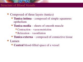
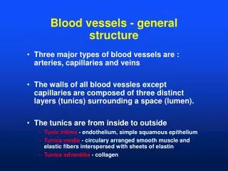
Arteries and Arterioles 1. Arteries are strong, elastic vessels adapted for carrying high-pressure blood. 2. The inside space of an artery through which blood flows is the lumen. • The wall of an artery has 3 layers. From the inside to the outside they are: • Tunica interna (endothelium)- Elastic tissue • Tunica media- Smooth muscle and elastic tissue • Tunica externa (adventitia)- Elastic tissue • The structure of an artery helps it propel blood onward while the ventricles are relaxing. • Arterioles are very small (almost microscopic) arteries
Arteries and Arterioles continued.. 6. When the sympathetic nervous system (fight or flight) is active, the smooth muscle of arteries contract, which narrows the lumen (less blood flow). This is called vasoconstriction. When the parasympathetic nervous system is active, the smooth muscle relaxes, which increases the lumen diameter. This is called vasodilation. *When would vasoconstriction occur? Vasodilation?
Tunica Interna Tunica Media Tunica Externa Artery – Cross section
Capillaries • Capillaries are the smallest vessels, consisting only of one cell layer through which substances are exchanged with tissue cells. Consist only of the tunica intima. • The number of capillaries throughout the body varies, depending on metabolic activity. Tissues with high metabolic requirements include the muscles, liver, kidneys and nervous system. 3. Because they are so numerous, blood flows more slowly through them which helps with substance exchange. 4. Precapillary sphincters (rings of smooth muscle) regulate the flow of blood into capillaries.
Exchanges in Capillaries 1. At the arteriole end of a capillary, the blood pressure is high, which forces oxygen, water and nutrients out of the capillary. This is called filtration. 2. At the venule end of a capillary, the blood pressure is lower, which causes most water and solutes that were forced out before to diffuse back into the capillary. This is called reabsorption. About 85% of the fluid filtered is reabsorbed. The carbon dioxide from the surrounding cells gets absorbed as well.
Filtration Reabsorption
Venules and Veins 1. Venules leading from capillaries merge to form veins that return blood to the heart. • Veins have the same three layers as arteries, but are much thinner. They do not carry the same high pressure blood. • Veins have flap-like valves inside to prevent backflow of blood. 4. Veins use the milking action of skeletal muscles to move blood because they are not lined with the same amount of smooth muscle as arteries.
Varicose Veins • In people with weak vein valves, gravity forces blood backwards through the valve. This increases venous blood pressure, which pushes the vein’s wall outward. After this repeatedly happens, the walls lose their elasticity and become stretched and flabby. This is called varicose veins.
Main Differences Between Blood Vessels • Walls of arteries are the thickest and their smooth muscle helps move blood • Lumens of veins are larger because walls are thinner • Larger veins have valves to prevent backflow • Skeletal muscle “milks” blood in veins toward the heart • Walls of capillaries are only one cell layer thick to allow for exchanges between blood and tissue
Blood Pressure • Blood pressure is the pressure exerted by blood on the walls of a blood vessel. It is highest in the aorta and large arteries branching off of the aorta. • Blood pressure is affected by the distance from the left ventricle , the total volume of blood, vasoconstriction/vasodilationandblood viscosity. • Pressure in an artery is approximately 93 mm Hg, in a capillary is 35 mm Hg and in a venule is 16 mm Hg. • Loss of blood would lower bp as water retention would raise bp. • Vasoconstriction raises bp as vasodilation lowers bp. • Blood viscosity is “thickness” of blood. The greater the # of blood cells and solutes, the greater the bp.
Systolic/Diastolic Pressure A healthy blood pressure reading is 120 mm Hg/80 mm Hg. • Systolic Blood Pressure is the top number of a blood pressure reading (120). This is the pressure that the blood is putting on the arteries as the ventricles contract, forcing blood out of the heart. • Diastolic Blood Pressure is the bottom number of a blood pressure reading (80). This is the pressure that the blood is putting on the arteries as the ventricles relax. • The surge of blood that occurs with ventricular contraction can be felt at certain points in the body as a pulse. The pulse rate normally is about 70-80 bpm.
Pulse • Pulse - Pressure wave of blood • Monitored at “pressure points” in arteries where pulse is easily felt • Pulse averages 70–76 beats per minute at rest
Variations in Blood Pressure • Normal human range is variable: • Normal • 140–110 mm Hg systolic • 80–70 mm Hg diastolic • Hypotension • Low systolic (below 110 mm HG) • Often associated with illness • Hypertension • High systolic (above 140 mm HG) • Can be dangerous if it is chronic
What helps maintain normal blood pressure? • Baroreceptors- Pressure receptor nerve cells which are located in the aorta and other large arteries and send messages to the brain. • Chemoreceptors- Measure the chemical composition (O2 and CO2) of blood. These cells send messages to alter respiration rate. • Hormones- Regulate blood flow and pressure • Renin, Adrenaline, ADH
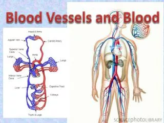
Systemic Circulation The systemic circuit includes the aorta and its branches leading to all body tissues as well as the system of veins returning blood to the right atrium.
1. External carotid-external to skull 2. Internal carotid- internal to skull 3. Common carotid- branches into external and internal 4. Vertebral- posterior brain 5. Brachiocephalic trunk-branches into right subclavian and right common carotid 6. Brachial-arm 7. Renal-kidney 8. Radial-radial side of forearm 9. Common iliac-branches into internal and external iliac 10. Internal iliac- pelvis 11. Subclavian- parts of brain, spinal cord, neck, chest, shoulder 12. Axillary- shoulder 13. Ulnar- ulna side of forearm 14. External iliac- lower limbs 15. Femoral- groin and thigh 16. Posterior tibial- posterior muscles and bones of leg and foot 17. Popliteal- knee and posterior leg muscles 18. Anterior tibial-anterior muscles and bones of leg and foot Major Arteries of Systemic CirculationAreas that they feed
Parts of the Aorta • The aorta has 4 parts: • Ascending aorta- Emerges from the left ventricle; where the coronary arteries branch to feed the heart. • Arch of the aorta- As the name implies, where the aorta “arches” to the left. The brachiocephalic trunk, left common carotid artery and left subclavian arteries branch off of this. • Thoracic aorta- The descending portion that is begins at the 4th/5th thoracic vertebrae and continues until the diaphragm opening. • Abdominal aorta- The descending portion between the diaphragm opening and the 4th lumbar vertebra.
1. Internal jugular- head 2. Vertebral- neck, cervical vertebrae 3. Brachiocephalic-dumps into the SVC 4. Brachial-forearm, elbow, and humerus 5. Renal- kidney 6. Radial- radius side of forearm 7. Common iliac- Pelvis and upper leg 8. Internal iliac- pelvis 9. Greater saphenous-longest in the body- femur region 10. Posterior tibial- back side of lower leg 11. Subclavian- arms, neck, thoracic wall 12. Axillary- arms and upper chest 13. Cephalic- lateral upper limbs 14. Hepatic-liver 15. Median cubital-elbow 16. Ulnar- ulna side of forearm 17. Femoral- thigh 18. Popliteal- knee 19. Small saphenous-Foot and posterior leg 20. Anterior tibial-anterior portion of leg, ankle and knee Major Veins of Systemic CirculationAreas that they drain
Common Blood Vessel Disorders • Hypertension- High blood pressure • Systolic greater than 140 mm Hg and Diastolic greater than 90 mm Hg • Effective treatments include: lose weight, limit alcohol intake, exercise, reduce sodium intake, don’t smoke and manage stress. • Aneurysm- A thin, weakened section of the wall of an artery or a vein. If it bursts, massive hemorrhaging could result. • Aneurysm video • Endovascular aneurysm repair