Download
1 / 29
330 likes | 538 Vues
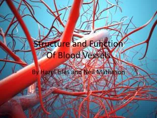
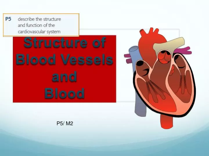
Structure of Blood Vessels and Blood. P5/ M2. The Heart. Muscular pump, about the size of a clenched fist Made up of a special muscle called Myocardium This can contract continuously without getting tired Main purpose is to drive blood through the arteries

E N D
The Heart • Muscular pump, about the size of a clenched fist • Made up of a special muscle called Myocardium • This can contract continuously without getting tired • Main purpose is to drive blood through the arteries • This delivers blood to the working muscles and other tissues.
Task… Find a picture of a blank heart (that is suitable to annotate) and upload it to a blank document. • Then do some light background research on the different labels of the heart including… • Structure of the cardiovascular system- heart: Atria Ventricles Bicuspid valve and tricuspid valve Aortic valveand pulmonary valve Aortaand vena cava – superior and inferior Pulmonary vein and pulmonary artery
The Heart continued • The Heart sits in a twin layered sac known as the Pericardium • Filled with pericardial fluid • Prevents friction as your heart beats.
The Heart continued • The heart wall has 3 layers • Epicardium (Outer) • Myocardium (middle layer and most of the heart wall) • Endocardium (The inner layer)
Atria/ Atrium • Upper chambers of the heart • Receive blood. • Right Atrium – receives deoxygenated blood from the body (Via the Vena Cava) • Left Atrium – receives oxygenated blood from the lungs (Via the pulmonary Vein) • Ventricles • Lower chambers of the heart • They have thicker walls and are stronger • Job is to pump the blood • Right Ventricle pumps blood to the lungs (pulmonary circulation) • Left ventricle pumps blood to the body (systemic circulation)
Valves • Tricuspid Valve • Bicuspid Valve • Also known as the mitral valve • Aortic Valve • Pulmonary Valve • All valves make sure that the blood flows in one direction, and there is no back flow • ChordaeTendineae • Cord like tendons that connect to the tricuspid and bicuspid valves • Ensure the valves stay the right way round and keep the blood flowing in the same direction.
Aorta and Vena Cava • The largest vein is the Vena Cava • Carries blood directly into the heart from the body • The largest Artery is the Aorta • Carries oxygenated blood directly out of the heart to the body tissues • Superior vena cava • brings blood from the upper body • Inferior Vena Cava • Brings blood from the lower body.
Pulmonary Circulation • Pulmonary Artery • Carries deoxygenated blood from the heart to the lungs. • It is the only artery that carries deoxygenated blood • Pulmonary Vein • Carries oxygenated blood from the lungs to the heart • It is the only vein that carries oxygenated blood.
The Heart = Double pump • To describe the flow of blood around the heart and the body, you will need to mention that the heart is made up of two pumps • Pulmonary circulation • Pumps blood to and from the lungs • Systemic circulation • Pumps blood around the body
Passage of blood flow • Blood flows into and out of the heart and around the body in one direction. • The heart is split into two distinct pumps by the Septum. • This makes sure that the oxygenated and deoxygenated blood don’t mix. • When describing the passage of blood flow around the heart, it is best to use a diagram and start on the right side of the heart.
The right side of the heart • Blood enters the heart (when it is relaxed) via the venacavae, • It goes into the rightatrium • The rightatrium contracts and the blood goes through the tricuspidvalve and into the right ventricle • The rightventricle contracts and the blood is pushed out of the heart through the semi lunar or pulmonaryvalve and into the pulmonaryartery • The pulmonaryartery carries the blood to the lungs • The heart relaxes and the valves close to stop back flow of the blood • In the lungs, the blood becomes oxygenated, and begins it’s journey back to the heart.
The Left side of the heart • The heart is relaxed, and this allows blood to enter the left side of the heart from the pulmonaryvein • It enters the leftatrium • The leftatrium contracts and pushes blood through the bicuspidvalve and into the leftventricle • The leftventricle has a very strong muscular wall and contracts very strongly. This closes the bicuspidvalve to prevent backflow, and pushes the blood through the aorticvalve and into the aorta. This is the largest artery and splits taking the blood to different areas of the body • The heart contracts and the aorticvalve closes, preventing back flow of the blood.
Terminology • Heart rate (H.R.) • The amount of times the heart beats in a minute. • Usually measured in beats per minute (b.p.m.) • Stroke Volume (S.V.) • The amount of blood leaving the left ventricle in one beat. • Normally measure in mililitres • Cardiac Output • The amount of blood leaving the heart in one minute • Normally measured in litres/minute • Cardiac Output = Stroke Volume x Heart Rate
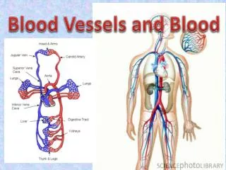
Structure of Blood Vessels and Blood
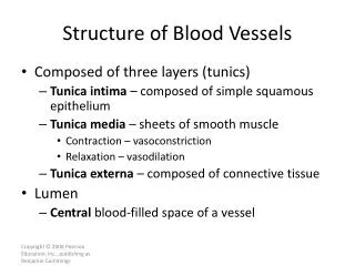
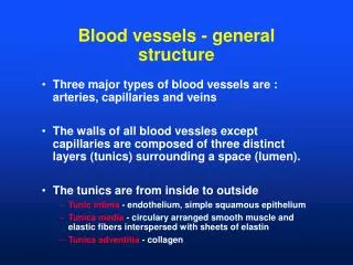
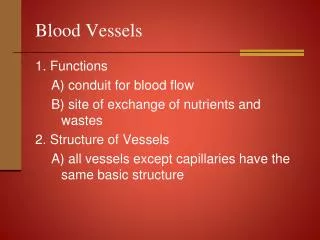
The Blood Vessels Introduction We have a variety of different vessels within the cardiovascular system to deliver and remove nutrients and waste products. Blood in the arteries is bright red, as it is carrying oxygen. It drops off the oxygen and picks up carbon dioxide as it moves through the capillaries. By the time it reaches the veins and venules it is a much darker blue/red colour.
5 main blood vessels • Carry blood away from the heart • Where gas exchange takes place • Carry blood back to the heart • Arteries • Arterioles • Capillaries • Venules • Veins
Arteries & Arterioles • Arteries are large blood vessels, that carry blood away from the heart. • Thick Elastic muscular walls • Artery walls contain elastic cartilage and smooth muscle, This allows the arteries walls to contract and relax to send blood to all parts of the body • This process is known as perstalsis, and is how smooth muscle contracts • Carry Oxygenated blood • Apart from the pulmonary artery, which carries deoxygenated blood to the lungs to get Oxygen
Arteries & Arterioles • Small round lumen • Operate under high pressure • Arteries don’t contain valves, as the blood is moving quickly under high pressure, so there is no chance of backflow. • Aorta • Main artery leaving the heart • It soon splits into smaller vessels – Arterioles • Arterioles deliver the blood to the capillaries
Capillaries • Smallest blood vessels • Found in all cells of the body • Just one cell thick • Very thin walls • Allows oxygen and other nutrients to diffuse through the cell walls. • Blood flows very slowly through the capillaries so that this can happen • Effectively in the capillaries, the blood unloads the Oxygen and picks up carbon dioxide and lactic acid (the waste products of metabolism)
Veins & Venules • The blood feeds from the capillaries back to the venules and then the veins. • Larger oval lumen • Means blood flows at lower speed and pressure. • Thinner and less muscular than arteries • Have some smooth muscle • Contracts to help sent the blood back to the heart
Veins & Venules • Carry blood back to the heart • Generally working against gravity • Particularly blood that is going back to the heart from the legs or arms, as it is below the heart. • Valves • To prevent the blood from flowing back once the smooth muscle relaxes. • Prevents pooling, particularly in the legs • Deoxygenated blood • Apart from pulmonary vein which carries oxygenated blood from the lungs back to the heart.
Task… • You are going to outline the function and characteristics of each of the 5 types of Blood Vessels that serve the circulatory system… • Including: • Physical characteristics that classify each blood vessel. • What their function is within the CIRCULATORY system? • What do they transport? Followed by a description/ explanation of the composition of BLOOD
Blood It is the medium in which all the cells are carried to transport nutrients and Oxygen (O2) to the cells of the body. It carries: - Oxygen, Glucose, Proteins, Fats, Vitamins, Hormones, Enzymes, Platelets, Carbon Dioxide and Electrolytes. Plasma: straw coloured liquid that all solids are carried within. • Made up of 4 components: • Red blood cells • White blood cells • Platelets • Plasma
Red Blood Cells Red blood cells make up 99% of the population of the blood cells in the body They are RED in colour due to the presence of a protein called HAEMOGLOBIN and absolutely loves Oxygen (massive attraction!) Making RED blood cells soul purpose to transport OXYGEN!
WhiteBlood Cells They are colourless and transparent and fewer in number to red blood cells (1:700) The role of White blood cells is to fight infection as they are part of the immune system. They destroy bacteria and other dangerous organisms… thus fighting potential infection.
Platelets They become sticky when in contact with air to form the initial stage of repair to the damaged tissue… They act by stopping blood loss through clotting Yet platelets need a substance called x-Factor 8 to enable them to become active and do their job/clot.
Describe the Structureand Functionof the cardiovascular system Examinethe cardiovascular system and explain how it works and how each part of the system is designed to meet its function Structure of the cardiovascular system (which you have to find pictures/ label and describe): Heart: Atria, Ventricles, Bicuspid valve, Tricuspid valve, Aortic valve, Pulmonary valve, Aorta, Vena cava – (superior and inferior), Pulmonary vein, Pulmonary artery Bloodvessels: Arteries, Arterioles, Capillaries, Veins, Venuoles Function of the cardiovascular system: Delivery of oxygen and nutrients Removal of waste products Thermoregulation(vasodilation and vasoconstriction of vessels); Function of blood (oxygen transport, clotting, fighting infection)