Download
1 / 80
800 likes | 1.05k Vues
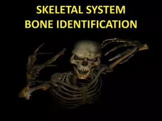
The Functions of the Skeletal System Bone Classification Bone Structure Bone Formation and Development Fractures: Bone Repair Exercise, Nutrition, Hormones, and Bone Tissue Calcium Homeostasis: Interactions of the Skeletal System and Other Organ Systems. SKELETAL SYSTEM.

E N D
The Functions of the Skeletal System • Bone Classification • Bone Structure • Bone Formation and Development • Fractures: Bone Repair • Exercise, Nutrition, Hormones, and Bone Tissue • Calcium Homeostasis: Interactions of the Skeletal System and Other Organ Systems
I’m all about the osseous tissues. That’s why I play the Trom-bone.
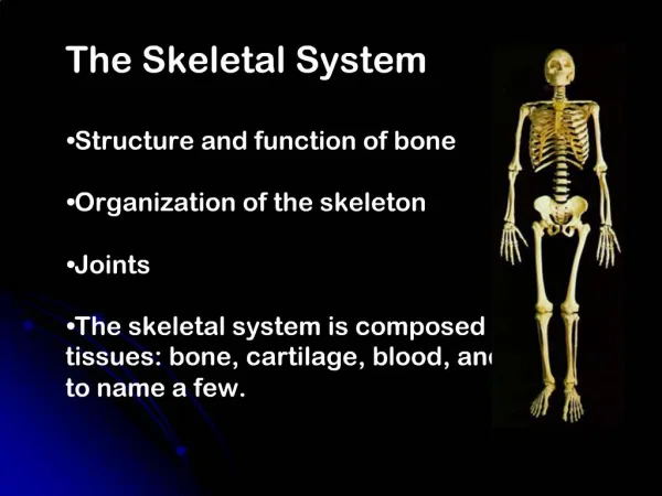
Osseous Tissue and the Skeletal Structure The skeletal system includes: Bones of the skeleton and associated cartilages Ligaments and other connective tissues that stabilize and/or connect them
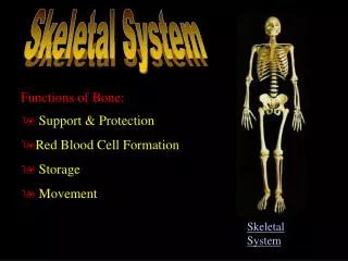
Primary Functions • Framework: support the body’s muscle fat, and skin (soft tissues). • Protection: Surround vital organs to protect them Examples • Skull that surrounds brain • Ribs that protect heart and lungs. • Levers: attach to muscles to help provide movement • Produce blood cells: produce red and white blood cells and platelets. • Storage: store most of body’s calcium, supply blood
Structure of bone • Matrix of Bone: 1/3 of bone weight is collagen fibers 2/3 of bone weight is calcium phosphate -Combination of the two provides strong bones which are somewhat flexible and resistant to shattering.
Terms • chondro refers to cartilage • chondrocyte • endochondral • perichondrium • osteo refers to bone • osteogenesis • osteocyte • periostium • blast refers to precursor cell or one that produces something • osteoblast • cyte refers to cell • osteocyte
Osteocytes= mature bone cells • recycle the calcium salts in the matrix around them • participate in the repair of damaged bone
Osteoblasts= responsible for the production of new bone. (osteogenesis) • Elevated local concentrations of calcium phosphate favorable for calcification
Osteoclasts= giant cells with 50 or more nuclei. • Derived from circulating monocytes (phoagocytic white blood cells) • Important in the regulation of calcium and phosphate concentrations in body fluids Osteoclast dissolving bone
Osteoprogenitor= mesenchymal cells that maintain populations of osteoblasts and play an important role in fracture repair
Long Bones- A long bone is one that is cylindrical in shape, being longer than it is wide. Long bones function as levers; they move when muscles contract. • Examples: humerus, ulna, radius, femur, tibia, fibula). • Keep in mind, however, that the term describes the shape of a bone, not its size.
Flat Bones- typically thin and often curved. Serve as points of attachment for muscles and often protect internal organs. • Examples include the cranial (skull) bones, the scapulae (shoulder blades), the sternum (breastbone), and the ribs. Flat bones
Short Bones- Short bones are approximately equal in length, width, and thickness • Examples: • The bones in the wrist and ankle
Irregular Bones- Bones that do not fall into the category of long, short, or flat are considered irregular bones. Examples: • The vertebrae and some of the skull bones are irregular.
Sesamoid bones- are small and round, and are located in tendons. • Examples: • patellae
Axial Skeleton (Anterior) • Creates the central axis of the body • Includes: • Skull • Thorax • Vertebral column • Contains 80 bones (~40% of skeleton)
Skull, Pelvis, and Vertebral columns are STRUCTURES NOT BONES!
Appendicular Skeleton 126 bones The bones of the limbs The pectoral girdle The pelvic girdle
Vertebral Column “Backbone” Central axis of skeleton 5 regions: Cervical vertebrae (C1-C7) Thoracic vertebrae (T1-T12) Lumbar vertebrae (L1-L5) Sacral (S) Coccygeal bone (CO) Different regions have special characteristics however all have common features *NOTE THE vertebrae numbering/identification! **HINT! HINT!**
Ribs & Costal Cartilages 12 pairs (24 total) One pair articulated with each thoracic vertebrae 7 True ribs – (T1-T7) Attach to sternum via cartilage (vertebrosternal ribs) 5 False ribs – (T8-T12) Do not directly attach to sternum (vertebrocondialribs) 2 Floating ribs – (T11-T12) SUB-TYPE OF FALSE RIB Not attached to sternum at all (vertebral ribs)
Upper Limb: Wrist & Hand Wrist – region between forearm and hand 8 carpals Hand – attached to carpals 5 metacarpals 5 digits Numbered 1-5 starting with thumb 3 phalanges per finger (2 on thumb) Proximal; Medial; Distal
Sutures- Immovable joints which tie bones firmly together with denser fibrous connective tissue
Sutures Lamboidal (λ)- Joins Parietal to Occipital Coronal(crown)-Joins Parietal to frontal Sagittal- joins the two parietal plates Squamosal (scale-like)- Joins temporal to occipital and parietal
Bone Markings • Bulges, depressions, and holes that serve as sites of attachment for muscles, ligaments, and tendons, joint surfaces, conduits for blood vessels and nerves.
Long Bone Flat Bone
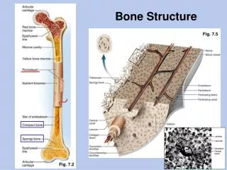
Compact Bone • Osteon= the functional unit of mature compact bone. (aka Haversian systems/canals) • Within osteon,osteocytes are arranged in concentric layers around the central canal. • Central Canals run parallel to the surface of the bone, contain one or more blood vessels.
Isolated osteon: • Nutrients diffuse from vessels in central canal • Alternating direction of collagen fibers increases resistance to twisting forces
Spongy Bone • Matrix of spongy bone: • Forms supportive mesh called trabeculae • Seem to align along stress lines • Has no osteonsor blood vessels. Nutrients reach osteocytes by diffusion
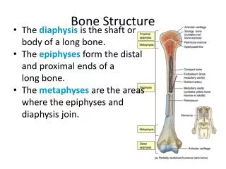
Diaphysis is the tubular shaft that runs between the proximal and distal ends of the bone • Medullary cavity- The hollow region in the diaphysis, which is filled with yellow marrow. The walls of the diaphysis are composed of dense and hard compact bone.
Epiphysis (plural = epiphyses)- The wider section at each end of the bone, filled with spongy bone. • Red marrow fills the spaces in the spongy bone.
Epiphyseal plate (growth plate)-narrow area where the epiphysis meets the diaphysis .Made of a layer of hyaline (transparent) cartilage in a growing bone. • When the bone stops growing in early adulthood (approximately 18–21 years), the cartilage is replaced by osseous tissue and the epiphyseal plate becomes an epiphyseal line.
The outer surface of the bone is covered with a fibrous membrane called the periosteum (peri- = “around” or “surrounding”). The periosteum contains blood vessels, nerves, and lymphatic vessels that nourish compact bone. WARNING NEXT SLIDE IS GRAPHIC
The medullary cavity has a delicate membranous lining called the endosteumwherebone growth, repair, and remodeling occur. • (endo- = “inside”; oste- = “bone”),
Tendons and ligaments also attach to bones at the periosteum. The periosteum covers the entire outer surface except where the epiphyses meet other bones to form joints.