Download
1 / 1
10 likes | 118 Vues
Dr Alkhansaa A.E.Ali (MB BS, MRCOG PART 1) Dr Nada Gaafar Hassan (MB BS, MD) Dr Shahad M. Hussein; (MB BS, MD, Fellow SMSB, MRCOG, CME) SOBA University Hospital University of Khartoum, Khartoum, Sudan. OPTIONAL LOGO HERE. OPTIONAL LOGO HERE. INTRODUCTION. CASE REPORT. DISCUSSION.

E N D
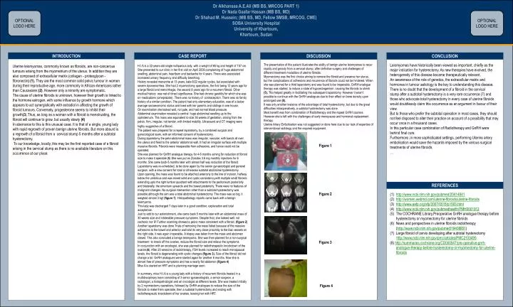
Dr Alkhansaa A.E.Ali (MB BS, MRCOG PART 1) Dr Nada Gaafar Hassan (MB BS, MD) Dr Shahad M. Hussein; (MB BS, MD, Fellow SMSB, MRCOG, CME) SOBA University Hospital University of Khartoum, Khartoum, Sudan OPTIONALLOGO HERE OPTIONALLOGO HERE INTRODUCTION CASE REPORT DISCUSSION CONCLUSION The presentation of this patient illustrates the ability of benign uterine leiomyomas to recur rapidly and grossly from a cervical stump, after definitive surgery and challenges of different treatment modalities of uterine fibroids. Myomectomy was the first choice aiming to remove the fibroid and preserve her uterus, but the complications of adhesions and recurrence of fibroids could not be hindered. When she was planned for a third laparotomy and was found to be inoperable, GnRH analogue therapy was started. to induce a state of hypoestrogenism, causing the fibroids to shrink (5). This helped greatly in facilitating the subsequent laparotomy. However it wasn’t possible to continue with the GnRH analogues due to their effect on bone density upon prolonged use (8). It was only another instance of the advantage of total hysterectomy, but due to the great difficulties intraoperatively, a subtotal hysterectomy was done. Most benefit was from combination of Radiotherapy and the repeat GnRH courses. However she is left with the challenges of early menopause and hormonal replacement therapy. Uterine Artery Embolisation was not suggested or done here due to our lack of expertise of interventional radiology and the required equipment. Figure 1 Figure 2 Figure 3 Figure 4 Leiomyomas have historically been viewed as important, chiefly as the major indication for hysterectomy. As new therapies have evolved, the heterogeneity of this disease became therapeutically relevant. An awareness of the role of genetics, the extracellular matrix and hormones in tumour aetiology is the key to understanding this disease. There is no doubt that the development of a fibroid in the cervical stump after a subtotal hysterectomy is a very rare occurrence (7) and those who advocate total hysterectomy in every case of uterine fibroids would doubtlessly claim this occurrence as an argument in favour of their view. But to those who prefer the subtotal operation in most cases, they should not feel disposed to alter their practice on account of a possibility that may occur once in a thousand cases. In this particular case combination of Radiotherapy and GnRH were behind final cure. Furthermore ,in more sophisticated settings, performing Uterine artery embolisation would save the hazards imposed by the various surgical treatments of uterine fibroids. … Uterine leiomyomas, commonly known as fibroids, are non-cancerous tumours arising from the myometrium of the uterus. In addition they are also composed of extracellular matrix (collagen - proteoglycan - fibronectin) (1). They are the most common solid pelvic tumour in women during their reproductive age, more commonly in African-Americans rather than Caucasians (2). However only a minority are symptomatic. The cause of uterine fibroids is unknown, however their growth is linked to the hormone estrogen, with some influence by growth hormone which appears to act synergistically with estradiol in affecting the growth of fibroid tumours. Conversely, progesterone seems to inhibit their growth(3).Thus, as long as a woman with a fibroid is menstruating, the fibroid will continue to grow, but usually slowly (4) In aberrance to this is this unusual case. It is that of a single, young lady with rapid regrowth of proven benign uterine fibroids. But more absurd is a regrowth of a fibroid from a cervical stump 5 months after a subtotal hysterectomy. To our knowledge, locally, this may be the first reported case of a fibroid arising in the cervical stump as there is no available literature on this occurrence at our place. H.I.A is a 32-years-old single nulliparous lady, with a weight of 65 kg and height of 157 cm. She presented to our clinic in her first visit on April 2009,complaining of huge abdominal swelling, abdominal pain, heartburn and backache for 4 years. There was associated increased urinary frequency and difficulty breathing. History revealed menarche at 13 years, kata 6/22 regular cycles, but associated with severe dysmenorrhea. She had 2 myomectomy operations, the first being 12 years ago for a large fibroid and menorrhagia, the second 8 years ago for a recurrent fibroid. Other medical history was not of direct significance. She had chronic gastritis for which she was on medications (omeprazole). There was no history of contraception. There was no family history of a similar condition. The patient had only elementary education, was of a below average socioeconomic status and lived with her parents and siblings in one house. On examination she looked well, not pale, and had normal blood pressure. Abdominal examination revealed a central huge abdominal swelling up to the xiphisternum. The mass was equivalent to size 36 weeks of gestation, arising from the pelvis, firm, irregular, not tender, with limited mobility. Ultrasound and CT imaging were highly suggestive of a fibroid. The patient was prepared for a repeat laparotomy, by a combined surgical and gynecological team, with an informed consent of hysterectomy. During laparotomy the pelvi-abdominal mass was irregular, vascular, with bands all over the uterus and fixed to the anterior abdominal wall. It had an irregular surface with multiple massive fibroids. Fibroids were inseparable from adhesions, and hence could not be operated. She was planned for GnRH analogue therapy for 4-6 months aiming for reduction of fibroid size to make it operable.(5) She was put on Zoladex 3.6 mg monthly injections for 6 months. She came back 6 months later with almost half way reduction of the fibroid. Laparotomy was re-scheduled, to be done again by the senior gynaecologist and general surgeon, with a new consent for total or otherwise subtotal abdominal hysterectomy. Upon opening, the mass was found to be attached anteriorly to the line of incision, halfway below the umbilicus and was mixed solid and cystic consistency,with multiple small fibroids extending upto the right lumbar quadrant with attachments to the peritoneum posteriorly and bilaterally, the omentum upwards and the bowel posteriorly. There were no features of malignant changes. No surgical intervention other than a subtotal hysterectomy was possible although the aim was a total abdominal hysterectomy. The mass was so big, it weighed almost 3 kg! (figure 1) Histopathology reports came back with a benign leiomyoma. This lady was discharged 7 days later in a good condition, explanation and total acceptance. Just to add to our astonishment, she came back 5 months later with an abdominal mass of 30 weeks size and intolerable pressure symptoms. Despite that, she looked well, not cachexic nor ill! Further scanning showed a pelvic mass consistent with a fibroid. (figure 2) Another laparotomy was done.Trials of removing the mass failed because of the massive adhesions to the bowel and anterior wall and its very close proximity to the iliac vessels on the right side, it was again inoperable. A biopsy was taken from the mass and abdomen closed. This also concluded a benign leiomyoma. She was then planned for a non-surgical treatment to knock off the ovaries, reduce the fibroid size and relieve the symptoms. In conjunction with an oncologist, she was planned for radiotherapeutic knockdown of the ovaries(6). After 20 sessions of radiotherapy, FSH levels increased to reach menopausal levels, the fibroid is degenerating with cystic changes (figure 3). Size of the fibroid did not change a lot. GnRH analogues were started again for another 6 months. Now she is almost free of pressure symptoms and has a nearly flat abdomen (figure 4) Miss A is started on HRT and is planning marriage soon. In summary, miss H.I.A is a young lady with a history of recurrent fibroids treated in a multidisciplinary team consisting of 3 senior gynaecologists, a senior surgeon, a radiologist, a histopathologist and an oncologist at different levels. She was treated initially by 2 myomectomy operations, followed by GnRH analogues to reduce the size of the fibroids to make them operable, then a subtotal hysterectomy and ending with radiotherapeutic knockdown of her ovaries, leaving her with HRT. REFERENCES • http://www.ncbi.nlm.nih.gov/pubmed/20414841 • http://women.webmd.com/uterine-fibroids/uterine-fibroids • http://www.aafp.org/afp/2007/0515/p1503.html • http://www.ncbi.nlm.nih.gov/pubmedhealth/PMH0001912 • The COCHRANE Library,Preoperative GnRH analogue therapy before hysterectomy or myomectomy for uterine fibroids • News and perspectives in uterine fibroids radiotherapy (http://www.ncbi.nlm.nih.gov/pubmed/19408851) • Large fibroid of cervix developing after subtotal hysterectomy • http://www.ncbi.nlm.nih.gov/pmc/articles/PMC2103495 • (8) http://summaries.cochrane.org/CD000547/pre-operative-gnrh-analogue-therapy-before-hysterectomy-or-myomectomy-for-uterine-fibroids