Download
1 / 18
180 likes | 378 Vues
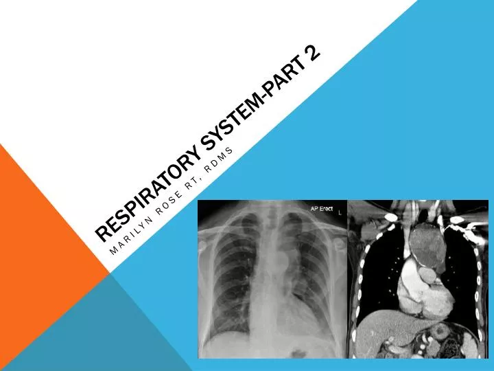
Respiratory system-part 2 . Marilyn Rose RT, RDMS. Outline Part 2. Neoplasms Vascular Diseases Miscellaneous Lung Disorders Disorders of the Pleura Mediastinal Masses Disorders of diaphragm. Neoplasm- benign. Solitary pulmonary nodule asymptomatic Incidental finding on CXR

E N D
Respiratory system-part 2 Marilyn Rose RT, RDMS
Outline Part 2 • Neoplasms • Vascular Diseases • Miscellaneous Lung Disorders • Disorders of the Pleura • Mediastinal Masses • Disorders of diaphragm
Neoplasm- benign • Solitary pulmonary nodule • asymptomatic • Incidental finding on CXR • Solitary nodule • Benign granuloma vs malignant mets /carcinoma • <30- minimal risk of Ca- <1% • 30-45 medium risk at 15% • >50- moderate risk – 50% • Radiograhic • Benign. • Central dense popcorn ca++ • Absence of growth on serial chest images over 2 years • Smooth/ sharp margins not spiculated/ ill-defined contour The enhancing rim: a new sign of a benign pulmonary nodule
Neoplasm- low grade • Bronchial adenoma • Low grade malignancy- 1% of all neoplasms • Equal in men and women • Appear in younger age groups • Hemoptysis and recurring pneumonia are common symptoms • Radiographic • Occur centrally in major segmental bronchi • Cause obstruction • Peripheral atelectasis/ pneumonitis due to bronchial obstruction
Neoplasm- malignant • Bronchogenic carcinoma- lung cancer • Primary cancer of the lung (mucosa) • squamous cell- arise in central bronchi, narrow lumen • adenocarcinoma= periphery • Bronchiolar- non-small cell- least common • Due to smoking and inhalation of carcinogens • Radiographic • Broad spectrum of radiographic abnormalities • Airway obstruction can cause atelectasis- • Pneumonia distal to obstructed bronchus • Absence of air bronchogram • Cavitation with a central necrosis- resemble abscess
Pulmonary Metastases • 1/3 of cancer patients develop pulmmets • Hematogenous or lymphatic spread • Most common from: MSK sarcoma, myeloma, breast Ca, urogenital, thyroid, colon • Radiographic: • Usually multiple, well cribumscribed, round nodules throughout the lungs • Thyroid= snowstorm of mets • Solitary- occur in 25%- may be indistinguishable from primary lesions or benign granuloma
Vascular disease • Pulmonary embolism • Potentially fatal • Most common path involving lungs of hospital patients • 80% have no symptoms • >95% emboli arise from thrombosis • develop in deep venous system • Rt side of heart • Most occlusions occur in the lower lobes • Consequence depends on size of emboli • Radiographic • Normal CXR • High resolution CT- base of heart to the top of the arch • Look for a filling defect or abrupt cutoff= complete obstruction
Vascular dz • Septic embolism • Shower of bacteria • Heart- bacterial endocarditis • Peripheral vein- septic thrombophlebitis • Hisotry of IV drug abuse • Radiographic • Almost always multiple • ill-defined • Cavitation frequently develop
Vascular dz • Pulmonary Arteriovenous Fistula • Abnormal vascular communication from a pumonary artery to a pulmonary vein • Large or multiple fistulas can cause shunting of bloon= cyanosis • Radiographic • Round or oval lobulated soft tissue mass • Lower lobe • Finding= feeding artery and draining vein (angio)
Miscellaneous Lung Disorders • Atelectasis • Condition where there is diminished air within the lung • associated with reduced lung volume • Result of bronchial obstruction • Neoplasm • Foreign body • Mucous plug ( can be an irritant from anesthesia) • Compression of lung from pneumothorax, fluid, tumor, abscess, emphysematous bulla • Iatrogenic= improper placement of ET tube- collapse left lung • Radiographic • Increase density by airless lung • Elevation of ipsilateralhemidiaphragm
Miscellaneous • ARDS • Adult respiratory distress syndrome • Variety of medical and surgical disorders • No major underlying disease • “ shock lung” • Pulmonary infection, aspiration, inhalation of toxins, drug overdose • Severe hypoxemia • Radiograohic • Patchy, ill defined • Consolidtion • Heart size remains normal
Miscellaneous • Foreign Body • Aspiration of solid foreign bodies into tracheobronchial tree • Almost always in young children • Compete obstruction leads to reabsorption of trapped air, alveolar collapse and atelectasis • Volume loss can cause heart to shift to affected side • Elevated ipsilateral diaphragm • Narrow intercostal spaces
Miscillaneous • Mediastinal Emphysema (Pneumomediastinum) • Air within mediastinal space • Spontaneous • Chest trauma- perf esophagus or tracheobronchial tree • Radiographic • Lateral displacement of mediastinal pleura, lateral= air posterior to sternum • Subcutaneous emphysema • Penetrating or blunt injury disrupting lung and parietal pleura • Forces air into tissue of chest wall • When palpating may feel crepitation (crackling) • Radiographic • Bizarre • Streaks of lucency outlining muscles
Disorders of the pleura • Pneumothorax • Presence of air in the pleural cavity • Partial or complete collapse of the lung • Traumatic, iatrogenic, hyaline membrane disease • Compress the lung and causes it to collapse • Radiographic • Hyperlucent area • All pulmonary markings are absent
Pleura • Pleural Effusion • Accumulation of fluid in pleural space • Nonspecific finding • Caused by a wide variety: CHF, PE, infection, neoplasm, result of abddzrecent OR, pancreatitis, subphrenic abscess • Radiographic • Blunting of costophrenic angles • Best in lateral CXR- posteriorly
Pleura • Empyema • Infected liquid or frank puss in plural space • Radiographic • Initially indistinguishable from plural effusion • It becomes loculated • Air-fluid level develops
Mediastinal masses • Anterior • thymoma, teratoma, thyroid mass, lipoma lymphoma • Middle • Lymph node disorders- lumphoma, mets, bronchogenic cyst • Posterior • Neurogenic tumor/ cyst, aneurysm of descending aorta • Most have no symptoms and are often found on routine CXR • Chest pain, dyspnea are caused by compression or invasion • Laryngeal nerve (hoarseness) and SVC syndrome= malignancy
Disorders of the diaphragm • Diaphragmatic Paralysis • Elevation of one or both leaves of the diaphragm • Interfere with phrenic nerve- surgical transection • Paradoxical movement of diaphragm ( “sniff” test or ULTRASOUND) • Eventration • Rare congenital abn where on hemidiaphragm is poorly developed/ weak • More frequent on LEFT • Bulging or elevation of diaphragm • rule out diaphragmatic hernia- abdominal contents will be displaced into chest • Elevated diaphragm…other • Ascites, obesity, pregnancy- condition where intra-abdominal volume is increased.