Download
1 / 15
150 likes | 310 Vues
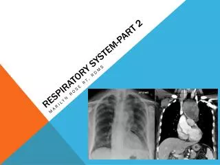
The Respiratory System 2. The respiratory system consists of an airway and lungs. T he airway conducts air into the lungs Breath of air passes in sequence : Nostrils, nasal cavities, pharynx, larynx, trachea, bronchi, bronchioles, and alveoli. The lower respiratory tract.

E N D
The respiratorysystemconsists of an airway and lungs • The airway conducts air into the lungs • Breath of air passes in sequence: • Nostrils, • nasal cavities, • pharynx, • larynx, • trachea, • bronchi, • bronchioles, • and alveoli
The lowerrespiratory tract • Trachea = windpipe • Main bronchi • Lobarbronchi • Lungs • Bronchioles • - Alveolarsacs • - Alveoli
The trachea = windpipe • Tube: • inner 20-25 mm indiameter • 10-16 cminlength • 15 – 20 incomplete C-shaped cartilaginous rings • The cartilaginous rings are incomplete because this allows the trachea to collapse slightly to allow food to pass down the esophagus • Posterior of trachea: smooth muscle and connective tissue trachea
Air passageway • Trachea two main bronchi (Left + Right) • The right main bronchus is wider, shorter, and more vertical than the left main bronchus • L main bronchus twolobar bronchi • R main bronchus three lobar bronchi • Lobar bronchi primary bronchioles • primary bronchioles alveolar ducts • alveolar ducts alveolar sacs alveoli
1: L main bronchus • 2: R main bronchus • 3: twolobarbronchi • 4: threelobar bronchi • 5: primary bronchioles • alveolarducts • 6: alveolarsacs • alveoli 1 2 3 4 5 6 6
The Lung • Every time you breath in and out your lungs are hard at work • Lungs are surrounded by the rib cage, on sit on diaphragm • The respiratory zone is the site of gas exchange with blood • Color: pink, after years becomes grey http://www.youtube.com/watch?v=nJuFmyXeHkA /
Parts • Base: bottom • Apex: tip • Hilus: placewherevessels, bronchiarestepin and out thelung
The Lung • The human lungs are the organs of respiration in humans • Humans have two lungs, with the left being divided into two lobes and the right into three lobes • Together, the lungs contain approximately 1500 miles (2,400 km) of airways and 300 to 500 million alveoli, having a total surface area of about 70 m2 in adults (the same area as a tennis court)
The pleura • The lungs are surrounded by the pleurae, a serous membrane which folds back upon itself to form a two-layered, membrane structure. • The thin space between the two pleural layers is known as the pleural space; it normally contains a small amount of pleural fluid. • The outer pleura (parietal pleura) is attached to the chest wall. • The inner pleura (visceral pleura) covers the lungs and adjoining structures, i.e. blood vessels, bronchi and nerves.