Download
1 / 1
10 likes | 123 Vues
i. b. b. m. e. 6 th International Meeting December 10-13 th , 2003 Orlando, Florida. Institute of Biomaterials and Biomedical Engineering. University of Toronto, CANADA. Figure 1. Scanning electron micrograph of an empty PLGA-CaP scaffold viewed longitudinally. D. C. 11 Days.

E N D
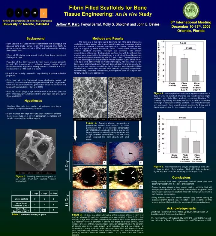
i b b m e 6th International Meeting December 10-13th, 2003 Orlando, Florida Institute of Biomaterials and Biomedical Engineering University of Toronto, CANADA Figure 1.Scanning electron micrograph of an empty PLGA-CaP scaffold viewed longitudinally. D C 11 Days 2 Days 5 Days Empty Scaffold 3 3 3 F E Fibrin Filled Scaffolds (Low T) 4 4 3 4 4 3 Fibrin Filled Scaffolds (High T) Table 1.Number of defects per group Fibrin Filled Scaffolds for Bone Tissue Engineering: An in vivo Study Jeffrey M. Karp, Feryal Sarraf, Molly S. Shoichet and John E. Davies • Background • Fibrin Sealants (FS) used clinically in combination with autologous and allogenic bone grafts (Tajima et al 1994, Sielewicz et al 1994), to deliver antibiotics (Marchettiet al 1994) and hydroxyapatite particles (Ferrari et al 1994). • Effects of FS during bony wound healing have been inconsistent (Schlaget al 1988). • Properties of the fibrin relevant to host tissue invasion generally ignored. FS investigated to enhance wound healing without modification to suit application (Huang2002 et al, Yamadaet al 2003, van Susanteet al 1999, Bach et al 2001). • Most FS are primarily designed to stop bleeding & provide adhesive properties. • Fibrin gels with thin fibers/small pores significantly reduce cell migration in vitro compared to fibrin gels with thick fibers/larger pores, which may be requirements of a gel structure critical for normal wound healing (Amraniet al 2001, Carr et al 1988). • Most FS contain (only) a high concentration of thrombin, (Jackson 2001) which results in a network with thin short fibers with small pores (Ryanet al 1999). Methods and Results The main goal of this work was to determine how filling bone engineering scaffolds with fibrin sealant effects bony wound healing and to examine whether the structural properties of the fibrin are important to consider. Tisseel VH was used as supplied by Baxter Bioscience Canada. To create fibrin matrices with different structural properties, fibrinogen was combined with different concentrations of thrombin. Biodegradable scaffolds filled with the different fibrin matrices were implanted into the distal femur of adult inbred Brown Norway rats and examined histomorphometrically after 2, 5 and 11 days. The results from the 5 day time point support many published in vitro cell migration studies where various cells types were demonstrated to migrate more rapidly into fibrin matrices with larger pores and thicker strands. This work demonstrates this phenomenon for the first time in vivo. However, results from the 11 day time point showed that filling scaffolds with commercially available fibrin sealants significantly delayed bony wound healing. Therefore, fibrin sealants, in their present state, are likely not ideal for bony wound healing applications. Figure 4. Histomorphometric analysis of reparative bone after 5 days in vivo. No statistical difference was found between empty scaffolds and fibrin filled scaffolds containing fibrin(low T). However, significantly less bone was found in defects containing fibrin(high T) compared to empty scaffolds. These results correlate with decrease in fibrin sealant amount between the 2 day and 5 day time points. (Low T ~ 62% reduction, high T ~ 34 %, p<0.06) Hypotheses 1. Scaffolds filled with fibrin sealant will enhance bone tissue invasion in comparison to empty scaffolds. 2. Fibrin matrices with large pores and thick strands will enhance bone tissue invasion in vivo in comparison to matrices with smaller pores and thinner fibrin strands. Figure 2. Scanning electron micrographs of polymerized Tisseel fibrin sealant. (A) Fibrin polymerized with a low thrombin concentration (1.75 NIH U/ml) contained thick fibrin strands with large pores compared to (B) fibrin polymerized with a high thrombin concentration (220 NIH U/ml), which contained thin fibrin strands with small pores. A B 5 Day Figure 5. Histomorphometric analysis of reparative bone after 11 days in vivo. Both scaffolds filled with fibrin contained significantly less bone then the empty scaffolds (p<0.003). • Conclusions • Filling Scaffolds with fibrin significantly reduces blood cells from becoming trapped within the pores of the scaffold. • During the early stages of bony wound healing, scaffolds filled with fibrin polymerized with a low thrombin concentration supported more bone invasion compared to scaffolds filled with fibrin polymerized with a high thrombin concentration. • Filling scaffolds with fibrin sealant delayed bony wound healing as evidenced after 11 days in vivo. Therefore, fibrin sealants in their present state are likely not ideal for bony wound healing applications. • Acknowledgements • Stuart Rae, Raisa Yakubovitch, Wanda Oprea, Dr. Tania Benatar, Dr. David Lickorish & Professor John Weisel. • This work was financially supported by an ORDCF awarded to JED and by a University of Toronto Doctoral Award and an OGS awarded to JMK. 11 Day Figure 3.(A) Bone was observed invading at the periphery of (low T) fibrin filled scaffolds whereas (B) much less reparative bone was identified in (high T) fibrin filled scaffolds. Large areas occupied by residual fibrin sealant (black arrow) were observed.(C) Reparative bone at periphery of defects was bright red-orange (arrows), which indicates presence of thick, mature collagen fibers. (D) Collagen fibers at the periphery of defects were green (white arrows), which indicates thin and less mature. In comparison to empty scaffolds, which were completely filled with bone(not shown) (E,F), bone was only observed at the periphery of both fibrin filled scaffolds groups. (A,B,E,F) Masson’s Trichrome stained sections. (C,D) Picro Sirus Red stained sections.