Download
1 / 31
320 likes | 364 Vues
Introduction to Surface Anatomy of upper & lower limbs. OBJECTIVES By the end of the lecture, students should be able to: Palpate and feel the bony the important prominences in the upper and the lower limbs. Palpate and feel the different muscles and muscular groups and tendons.

E N D
OBJECTIVES • By the end of the lecture, students should be able to: • Palpate and feel the bony the important prominences in the upper and the lower limbs. • Palpate and feel the different muscles and muscular groups and tendons. • Perform some movements to see the action of individual muscle or muscular groups in the upper and lower limbs. • Feel the pulsations of most of the arteries of the upper and lower limbs. • Locate the site of most of the superficial veins in the upper and lower limbs Prof. Saeed Abuel Makarem
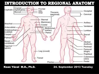
What is Surface Anatomy? • It is a branch of gross anatomy that examines shapes and markings on the surface of the body as they are related to deeper structures. • It is essential in locating and identifying anatomic structures prior to studying internal gross anatomy. • It helps to locate the affected organ / structure / region in disease process.
The clavicle is subcutaneous and can be palpated throughout its length. • Its sternal end projects little above the manubrium. • Between the 2 sternal ends of the 2 clavicles lies the jugular notch (suprasternal notch). • The acromial end of the clavicle can be palpated medial to the lateral border of the acromion, of the scapula. particularly when the shoulder is alternately raised and depressed. • The large vessels and nerves to the upper limb pass posterior to the convexity of the clavicle.
The coracoid process of scapula can be felt deeply below the lateral one third of the clavicle in the Deltopectoral GROOVE or clavipectoral triangle. • Theclavipectoral or the (Deltopectoral) triangleis the slightly depressed area just inferior to the lateral third of clavicle. • The clavipectoral triangle is bounded by: • Clavicle superiorly, • Deltoid laterally, and • Pectoralis major medially.
The lateral and posterior borders of the acromion meet to form the acromial angle. Inferior to the acromion, the deltoid muscleforms the rounded contour of the shoulder.
The greater tubercle of humeruscan be felt by deep palpation through the deltoid muscle, inferior to the acromion when the arm is by the side. • In this position, the greater tubercle is the most lateral bony point of the shoulder. • The shaft of the humerusmay be felt in different areas through the muscles surrounding it. • The medial and lateral epicondyles of the humerusare palpated on the medial & lateral sides of the elbow region.
The head of ulna forms a rounded subcutaneous prominence that can be easily seen and palpated on the medial side of the dorsal aspect of the wrist. • The pointed subcutaneous ulnar styloid process may be felt slightly distal to the ulnar head when the hand is supinated. • The olecranon and posterior border of the ulna lie subcutaneously and can be palpated easily. • When the elbow joint is extended, the tip of the olecranon process, the medial and the lateral epicondyles lie in a straight line. • When the elbow is flexed, the olecranon forms the apex of an equilateral triangle, of which the epicondyles form the angles at its base. • Fractures of any of these structures will disturb this arrangement.
The head of radius can be palpated and felt to rotate in the depression on the posterolateral aspect of the extended elbow, just distal to the lateral epicondyle of the humerus with supination and pronation. • The radial styloid process can be palpated on the lateral side of the wrist in the anatomical snuff box. • It is approximately 1 cm distal to that of the ulna. Prof. Saeed Abuel Makarem
The metacarpals, although they overlapped by the long extensor tendons of the fingers, they can be palpated on the dorsum of the hand. • The heads of the metacarpals form the knuckles of the hand. • Notice that the 3rd metacarpal head is the most prominent. • The dorsal aspects of the phalangescan be easily palpated. • The knuckles of the fingers are formed by the heads ofthe proximal and middle phalanges.
Axillary Folds • The anterior axillary fold is formed by the lower margin of the pectoralis major, and can be palpated by the finger. • This can be made by asking the patient to press his or her hand against the ipsilateral hip. • The posterior axillary fold is formed by the tendons of latissimus dorsi and teres major muscles • Axilla • The axilla should be examined with the forearm supported and the pectoral muscles relaxed. • When the arm by the side, the inferior part of the head of the humerus can be easily palpated through the floor of the axilla. • The pulsations of the axillary artery can be felt high up in the axilla, and around the artery the cords of the brachial plexus. • The medial wall of the axilla is formed by the upper ribs covered by the serratus anterior. • The lateral wall is formed by the coracobrachialis and biceps brachii and the bicipital groove.
The borders of the deltoidare visible when the arm is abducted against resistance. • The distal attachment of the deltoid can be palpated on the lateral surface of the humerus. • Biceps brachii & triceps brachii form bulge on the anterior and posterior surfaces of the arm. • The biceps tendon can be palpated in the cubital fossa, immediately lateral to the midline. • The triceps tendon can be palpated where it is attached to the olecranon process. • There are 2 grooves: Medial and lateral groovesseparate the bulges formed by the biceps and triceps. • The cephalic veinascends superiorly in the lateral groove and • The basilic veinascends in the medial groove.
The brachial artery can be felt pulsating deep to the medial border of the biceps. To stop bleeding by pressure on the artery in the upper half of the arm it is pushed laterally against the humerus. In the lower half it is pushed posteriorly. In the cubital fossa, it lies beneath the bicipital aponeurosis. At the level of the neck of the radius, it divides into radial and ulnar arteries. Brachial artery
CUBITAL FOSSA In the cubital fossa, try to locate: • Cephalic vein • Basilic veinand • Median cubital veinare clearly visible. • The median cubital vein connects the cephalic and the basilic veins . • It crosses over the bicipital aponeurosis. • It is the vein of choice for IV line, WHY?
DORSUM OF THE HAND The dorsal venous network: The network of superficial veins can be seen on the dorsum of the hand. The network drains upward into the cephalic vein laterally, and the basilic vein medially. The tendons of extensor digitorum, extensor indicis, and extensor digiti minimi can be seen and felt as you extends your fingers.
ANATOMICAL SNUFF BOX • In its proximal part the radial styloid process is palpable. • The scaphoid boneis also palpable in the distal part of the anatomical snuff box. It is a depression on the lateral aspect of the wrist joint which is accentuated when you extends your thumb. Boundaries • The snuff box is bounded : • Anteriorlyby 2 tendons: • Abductor pollicis longus • Extensor pollicis brevis • Posteriorlyby extensor pollicis longus
The Radial arterycan be drawn by a line extends from the midpoint of the cubital fossa to the base of the styloid process of radius. Radial Artery pulsation: Universally, its pulsations can easily be felt anterior to the distal third of radius. Here it lies just beneath the skin and fascia lateral to the tendon of flexor carpi radialis muscle
Also, the radial artery pulsation can be felt against the floor of the snuff box. • More superficially, the anatomical snuff box is crossed by • The cephalic vein. • The radial nerve.
Superficial Palmar Arterial Arch. The superficial palmar arterial arch is located in the central part of the palm and lies on a line drawn across the palm at the level of the distal border of the fully ex-tended thumb. Deep Palmar Arterial Arch. The deep palmar arterial arch is also located in the central part of the palm ( proximal to the superficial one), lies on a line drawn across the palm at the level of the proximal border of the fully extended thumb.
All of the following structures are palpable in the inguinal region: Symphysis pubis Body of pubis Pubic tubercle ASIS
The inguinal ligament extends between: The pubic tubercle and The ASIS. In the mid-inguinal point you can feel the pulsations of the femoral artery. The femoral vein lies on the medial side of the artery. While the femoral nerve lies lateral to the artery.
Midinguinal point: It is a point on the inguinal ligament midway between the symphysis pubis and the ASIS. The femoral artery is an important site for vascular access as a large number of arteriographic procedures are undertaken through its percutaneous puncture, (coronary angiography).
Femoral Triangle The femoral triangle can be seen as a depression below the fold of the groin in the upper part of the thigh. In a thin, muscular subject, the boundaries of the triangle can be identified when the thigh is flexed, abducted, and laterally rotated. The base of the triangle is formed by the inguinal ligament, the lateral border by the sartorius and the medial border by the adductor longus
The iliac crest is subcutaneous and can be palpated throughout its length, from the ASIS to the PSIS. The greater trochanter of the femur is also subcutaneous and can be palpated on the lateral aspect of the hip joint behind and distal to the ASIS.
KNEE REGION In front of the knee joint the patella and the ligamentum patellae can be easily palpated. The ligamentum patellae can be traced downward as it is attached to the tibial tuberosity. The condyles of the femur and tibia can be recognized on the sides of the knee and the joint line can be identified between them.
On the medial aspect of the knee Joint try to palpate: • Medial femoral condyle • Medial tibial condyle • The 3 tendons of • sartorius. • Gracilis • Semitendinosus. On the lateral aspect of the knee Joint try to palpate: Lateral femoral condyle Lateral tibial condyle Head of the fibula Neck of the fibula Tendon of biceps femoris.
In the back of the knee and leg try to palpate: The boundaries of the popliteal fossa. The pulsation of the popliteal artery which is deeply situated in the fossa.
On the anterior aspect of the leg and knee Joint and try to palpate: The patella. The tibial tuberosity. The anterior border of the tibia, (shine). The medial tibial condyle. The medial surface of the tibia. The medial malleolus. The lateral malleolus. On the dorsum of the foot try to palpate: The tuberosity of the 5th metatarsal The tubercle of navicular. The metatarsals.
On the dorsum of the foot try to palpate: The long extensor tendons: Tibialis anterior Extensor hallucis longus. Extensor digitorum longus. Peroneus tertius. Also, try to feel the pulsation of the dorsalis pedis artery. Between the tendons of extensor hallucis longus & extensor digitorum longus.
On the lateral aspect of the leg try to palpate: The tendons of peroneus longus and brevis. The tendon Achilles. The lateral malleolus. Prof. Saeed Abuel Makarem
On the Medial aspect of the ankle try to palpateand feel: The medial malleolus. The tendons of tibialis posterior The tendon of flexor digitorum longus. The posterior tibial artery The calcaneus.