Download
1 / 34
370 likes | 541 Vues
Retinal Bioengineering. http://www.eye-chip.com/ http://www.bostonretinalimplant.org/ http://www.eyesight.org/ http://www.amdcanada.com/ A lot of the material in the first part of this lecture is available online: http://webvision.med.utah.edu/index.html. Nov 13 th , 2006. outline.

E N D
Retinal Bioengineering http://www.eye-chip.com/ http://www.bostonretinalimplant.org/ http://www.eyesight.org/ http://www.amdcanada.com/ A lot of the material in the first part of this lecture is available online: http://webvision.med.utah.edu/index.html Nov 13th, 2006
outline • Intro: retina, eye, visual system. • Retinal structure and function. • Retinal diseases (rp, md, glaucoma, detachment) • Engineering contributions to retinal physiology • Implants: epi/subretinal • homework
How do we project light stimuli? Optics of the Human EyeBy David A. Atchison, George Smith http://thalamus.wustl.edu/course/bvis2.gif
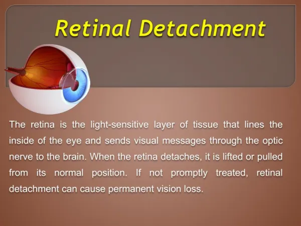
Anatomy of the eye http://webvision.med.utah.edu/sretina.html
Retinal pigment epithelium Providing life support for the photoreceptors is a layer of the retina called the retinal pigment epithelium, or RPE. The RPE is a single layer of retinal cells, densely packed with pigment granules, that is located between the photoreceptors and Bruch's Membrane. Each RPE cell is in contact with approximately 25 rods and/or cones. Rod and cone cells each shed approximately 100 discs (waste products) every day as part of their normal function, and this waste is cleared by the RPE. Over the course of a lifetime, 70 million discs may be cleared or "digested" by each RPE cell. One theory on the development of AMD is linked to a breakdown of this 'digestive' action. http://www.amdcanada.com/template.php?section=4&subSec=2d&content=4_2
Retinal layers: nomenclature indicates cell types and synaptic connections http://thalamus.wustl.edu/course/eyeret.html
They span from inner limiting membrane (ILM) all the way to ganglion cell layer (GCL). They are vertically positioned, like the bipolar cells. They form gap junctions in several species, and take care of the extracellular potassium (potassium buffer Muller glial cells: special glia cells, only in the retina. http://webvision.med.utah.edu/imageswv/FisherFig1.jpg
PhototransductionRods and cones: how much current per incident “photon” Pathways from rods and cones to ganglion cells are not the same, even though each single ganglion cell receives input from both rods and cones. Amacrine cells intermediate connections from rods to ganglion cells (through bipolar cells) http://webvision.med.utah.edu/imageswv/rodcoEM.jpeg
Humans have two kinds of light transducers: rods and cones. There are three basic kinds of cones, with different spectral sensitivities. When it’s dark, we “see” with rods. When it’s light, we “see” with cones. http://webvision.med.utah.edu/imageswv/spectra.jpeg
Differential sensitivity of rods and cones Rod = 0.7pA* 30 rods/bipolar light Cone = 0.033pA* 4 cones/bipolar * Result of the primate photoisomerization in the outer segments of rods and cones. Visual angle of common objects (degrees, deg) The sun or moon = 0.5 deg Thumbnail (at arm's length) = 1.5 deg Fist (at arm's length) = 8-10 deg
Photons absorbed by rods and cones rod cone Number of photons absorbed Light stimulus Time (s) Neural Engineering, Bin He. chap 13
Fovea, cell density • Fovea: cones packed together, form hexagonal pattern http://upload.wikimedia.org/wikipedia/en/2/2d/Retina-OCT800.png http://webvision.med.utah.edu/sretina.html#muller
How small can you see? Stimulus: black & white bars Our best sensors: cones in the fovea. Cone: one element. Packed densely: 2.5mm center to center (this is the element spacing). Distance between two lines: one cycle R = 300mm/degree x [1 cycle / (2 x element spacing)], Where R is the resolution in cycles of the grating per degree. What is the resolution of your eye?
Numbers… • Typical ambient luminance levels (in cd/m2): • Starlight: 0.001 • Moonlight: 0.1 • Indoor lighting: 100 • Sunlight: 10,000 • Maximum intensity of CRT monitors: 100 • One Troland (Td) of retinal illumination is produced when an eye with a pupil size of 1 mm2 looks at a surface whose luminance is 1 cd/m2. • Lens focal length: f(meters); lens power= 1/f (diopters). • Obs: Lux are units of illumination. Light intensity of 1 candela produces an illumination of 1 lux at 1 meter. • Blind spot: where the optic nerve • A X/Y vision means that the numerator person can see at X feet what a normal person can see at Y feet. Usual numbers are 20/20 (normal vision); 80/100 (means the patient has to be at 80 feet to see what a normal subject would see at 100 feet). Modified from http://webvision.med.utah.edu/
ON/OFF bipolar cells http://webvision.med.utah.edu/imageswv/bcfig1.jpg
On/off center bipolar cells Neural Engineering, Bin He. chap 13
Receptive fields: space mapping of light stimuli to ganglion cells http://webvision.med.utah.edu/imageswv/SK-SPOTS.JPG
Retinal vasculature Two sources of blood supply to the mammalian retina: (1) the central retinal artery (15-35% of the blood flow, supply to the inner retinal layers; (2) the choroidal blood vessels. (65-85% of the blood flow, supply to photoreceptors through the pigment epithelium). http://webvision.med.utah.edu/imageswv/FlorretBV.jpg
Retinal diseases: 15 10^6 blind or visually impaired in the U.S.A. • Retinitis pigmentosa • Macular degeneration • Glaucoma • Diabetic retinopathy • Vascular occlusive disease • Retinal detachment Most prevalent diseases. (4th: age-related cataract)
Less rods and cones Inner retinal layers “receive” too much O2 Less oxygen consumed vasoconstriction Permanent damage Retinitis pigmentosa • Affects one in 4k to 3k; • Characteristics: photoreceptor loss. (rods first, cones second); • Cause: more than 50 genetic defects in photoreceptor or pigment epithelium proteins.
Age-related macular degeneration • Prevalence: 1 in 100 (adults over 40y.o.) • Incidence: higher over 65 y.o. • Photoreceptor degeneration (similar to RP): incomplete digestion of outer segment disks (lipids and proteins) • Drusen → traffic jam between the choroid and the retina → prevents metabolites from being delivered → neovascularization (choroidal vessels proliferate and enter the retina). http://www.amdcanada.com/images/content/3_3_2_2_fig3.jpg
Vascular occlusive disease • Atheorsclerosis in arteries or veins (like a stroke in the retina) • No redundancy in circulation (remember theory of cell assemblies), so occlusion leads to scotoma. • if t>2h, then (damage = permanent) • Venous occlusion → hemorrhages → less damaging than arteries.
IOP control (outflow) Glaucoma • Prevalence: 0.8 in 100 (to 3 in 100 Caucasians) • Damaged ganglion cells due to elevated intraocular pressure (IOP). • Normal: 15mm Hg. Glaucoma: 22mm Hg. • High pressure compresses optic nerve, axonal transport is blocked, retrograde degeneration of ganglion cells. http://www.amdcanada.com/images/content/4_2_fig1.jpg
Electroretinogram http://webvision.med.utah.edu/imageswv/ERGFig3.jpg
Waves within the ERG http://webvision.med.utah.edu/imageswv/
Photoreceptor models: based on the ERG Neural Engineering, Bin He. chap 13
ERG analyses Neural Engineering, Bin He. chap 13
Engineering insights What can you do to help? Considerations on designing an implant: - do users (patients) want to be helped? - what do you think they’d answer as to what the most important specification of your implant/system would be?
Retinal implants www.bostonretinalimplant.org
NCSU Retina Prosthesis Team NCSU Open House • NCSU Staff • Dr. Wentai Liu, PhD • Mark Clements (PhD student) • Kasin Vichienchom (PhD student) • Primary tasks • Image acquisition (camera) • Image processing • Micro-stimulator design • WWW address • http://www.ece.ncsu.edu/retina • Mark Clements (PhD student) • Kasin Vichienchom (PhD student) • Chris DeMarco (PhD student)
Retinal transplant. Progenitor cells. http://www.bmc.riken.jp/~yagi/retina/