Download
1 / 73
1.08k likes | 2.69k Vues
OBSTRUCTIVE JAUNDICE. Presented by: MD. TANVIR MAJUMDER, Roll no.144 MD. MAHEDI HASAN, Roll no.150 MONIRUL HOQUE, Roll no.162 MD. SHAHADATH HOSSAIN, Roll no.163. CASE PRESENTATION ON: Obstructive jaundice. PARTICULARS OF THE PATIENT :.

E N D
OBSTRUCTIVE JAUNDICE Presented by: MD. TANVIR MAJUMDER, Roll no.144 MD. MAHEDI HASAN, Roll no.150 MONIRUL HOQUE, Roll no.162 MD. SHAHADATH HOSSAIN, Roll no.163
CASE PRESENTATION ON: • Obstructive jaundice
PARTICULARSOFTHEPATIENT : Name : Mr. Kala Mohon Das Age : 65 years Fathers name :Late Nagar Baul Das Sex : Male Marital status: Married Religion : Sanatan Occupation: Garment worker Address: Gangabari, Pathorghata, Kotowalli, Ctg Bed no. : 18 Ward no.: 25 Date of admission :7-4-2013; 7pm Date of examination :25-4-2013;8pm
THE PRESENTING COMPLAINTS: • 1. recurrent pain in the right upper abdomen for last 1 year. • 2. yellow discoloration of eyes and skin for the same duration.
HISTORY OF PRESENT ILLNESS: According to the patient’s statement his presenting complaints started 1 year back. He developed severe pain in right upper abdomen which is colicky in nature, intermittent, radiating to the back on the tip of the scapula. The pain aggravated by taking food and relieved by medication, which was episodic occurring at an interval of 1 or 2 months. He had moderate grade, intermittent fever which was associated with chills and rigor. He also complained of vomiting after taking any food. Vomitus was non projectile, whitish in colour and contained both digested and undigested food particles. He had yellow discolouration of eyes and urine for the same duration. His bowel habit is normal. He has no history of pale colouration of stool. He has itching all over the body. He also has no history of weight loss, cough, hemoptysis, haematemesis, melaena or bone pain.
HISTORY OF PAST ILLNESS: • Patient gave no history of DM, HTN, TB, bronchial asthma.
PERSONAL AND SOCIO-ECONOMIC HISTORY: • Patient is non-smoker, non-alcoholic. • He comes from a lower-middle class family. He lives on average diet and uses sanitary latrine. FAMILY HISTORY: None of his family members is known to be suffering from the same or any other diseases.
DRUG HISTORY : He used to take analgesics and antipyretics for the last 3 months but could not mention the name of the drugs. ALLERGIC HISTORY: • He is not allergic to any drug or any particular food. TRANSFUSION HISTORY: • Patient has no history of blood transfusion.
Appearance: Anxious • Body built: Average • Nutrition: Average • Co-operation: Cooperative • Decubitus: On choice • Anaemia: Absent • Jaundice: PRESENT (++) • Cyanosis: Absent • Clubbing: Absent • Edema: Absent • Dehydration: Absent • Pulse: 80 beats/minute • Blood pressure: 110/80 mm of Hg • Temperature: 100 degree F • Respiratory rate: 18 breaths/minute • Neck vein: Not engorged • Neck gland: Not enlarged • Peripheral lymph node: Not palpable • Hernial orifice: Intact
Abdomen examination • Abdomen is normal in shape. • Umbilicus is centrally placed, inverted, vertical slit. • No engorged vein. • No visible peristalsis. • No scar mark. • Hair distribution normal. • No visible pulsation. inspection
SUPERFICIAL PALPATION : • Hyperesthesia: absent • Tenderness: present in right hypochondriac region. • Temperature : raised • Muscle guard: absent DEEP PALPATION • Liver : not palpable • Gall Bladder: not palpable • Spleen: not palpable • Kidney: not palpable • Urinary bladder :not palpable. • Murphy’s sign: negative
Percussion • Note: Tympanic • Shifting dullness and fluid thrill absent
Auscultation • Shows no abnormality • Bowel sound present digital rectal examination
Cardiovuscular system: Peripheral pulses: All are present,symmetrical. Precordium: Apex beat: Left 5th ICS, 9 cm from midline Thrill: Absent Left parasternal heave: Absent Heart sound: Audible normally in all 4 areas Added sound: Absent
Respiratory system: Position of trachea and apex beat: Normal Chest expansion: Normal Percussion note: Resonant Breath sound: Normal Added sound: Absent
Nervous system Higher Mental function: Normal Cranial nerves: Intact Speech: Normal Signs of meningeal irritation: Absent Motor function: Intact Sensory function: Intact
SALIENT FEATURES: Mr. Kala Mohon Das, 65 years old, hailing from Kotowalli, Chittagong presented with the complaints of pain in the right upper abdomen for last 1 year, and yellow discoloration of eye and skin for the same duration. According to the patient’s statement, his presenting complaints started 1 years back when he developed pain in right upper abdomen which was severe, colicky in nature, intermittent, radiating to the back, aggravated by taking food and relieved after taking medication, which was episodic occurring at an interval of 1 or 2 months. He had moderate grade, intermittent fever which was associated with chills and rigor. He also complained of vomiting after taking any food. Vomitus was non projectile, whitish in colour and contained both digested and undigested food particles. He had yellow discolouration of eyes and urine for the same duration. His bowel habit is normal. He gives no history of pale colouration of stool. He has itching all over the body. He gave no history of weight loss, cough, chest pain, hemoptysis, haematemesis, melaena and bone pain. He has no history of blood transfusion. No member of his family is suffering from this disease. He is non-smoker, non-hypertensive, not diabetic.
On general examination, patient is anxious,co-operative,average built. He is icteric but not anaemic. Neck gland are not palpable, neck veins are not engorged, no ascites, no oedema is present. Hernial orifices are intact and all accessible peripheral lymph node are not palpable. His pulse, B.P., respiration was within normal limit and there was slight rise of body temperature. On abdominal examination, abdomen is normal in shape, umbilicus centrally placed,inverted,no scar mark, no engorged vein. Murphy’s sign is negative. There is tenderness in right hypochondriac region. Liver or other organs are not palpable. Bowel sound is present. Other systemic examination reveals no abnormality.
PROVISIONAL DIAGNOSIS: • OBSTRUCTIVE JAUNDICE due to choledocholithiasis
DIFFERENTIAL DIAGNOSIS: Obstructive jaundice due to: • carcinoma head of pancreas • Biliaryascariasis • Periampullary carcinoma • Cholangiocarcinoma
INVESTIGATIONS: 1.USG of whole abdomen with special attention to hepatobilliary system and pancreas: To visualize- -gall bladder -Liver -LN -Ascites 2.LIVER FUNCTION TEST: -serum bilirubin -SGOT -SGPT -Alkaline phosphatase -prothrombin time 3.FOR GENERAL ASSESSMENT: -CBC -urine R/E -RBS -serum creatinine -CXR -ECG
Confirmatory diagnosis: • Obstructive jaundice due to choledocholithiasis.
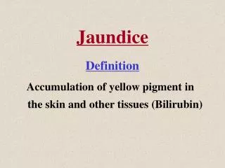
What is jaundice ? Jaundice may be defined as yellow discolouration of skin,sclera and mucous membrane due to an increase billirubin concentration in the body fluid above normal. Serum total bilirubin:(normal 0.3-1.2 mg/dl ) clinically detectable: >3.0 mg/dL
Pathophysiologic classification of Jaundice • Hemolytic Jaundice • HepatocellularJaundice • Obstructive Jaundice
OBSTRUCTIVE JAUNDICE: • Obstructive jaundice occurs due to mechanical obstruction of the common bile duct.
CAUSES OF OBSTRUCTIVE JAUNDICE: 1.IN THE LUMEN: -gall stone -round worm 2.IN THE WALL: -congenital atresia -traumatic stricture -cholangitis -tumour of the bile duct 3.OUTSIDE THE WALL: -carcinoma head of the pancreas -carcinoma of ampulla of vater -pancreatitis -enlarged LN at portahepatis due to metastasis.
EXAMINATION FINDINGS OF OBSTRUCTIVE JAUNDICE: B.CARCINOMA HEAD OF PANCREAS: 1.Palpable liver 2.palpable mass 3.splenomegaly 4.palpable gall bladder. 5.weight loss. A.CHOLEDOCHOLITHIASIS: 1.patient-toxic,ill-looking 2.jaundice-long standing produce a deep green hue. 3.dehydrated 4.skin scratch mark all over the body. 5.increased temperature. 6.no lymphadenopathy. 7.tenderness in right hypochondriac region. 8.gall bladder is not palpable. 9.liver may be enlarged.
EXAMINATION FINDINGS OF OBSTRUCTIVE JAUNDICE: D.CHOLANGIOCARCINOMA: 1.patient is cachectic, anaemic 2.ichteric,dehydrated 3.hepatomegaly 4.palpable gall bladder-if lesion below cystic duct 5.ascitis-in advanced cases. C.PERIAMPULLARY CARCINOMA: 1.Fluctuating jaundice 2.palpable gall bladder 3.ascitis. 4.weight loss 5.palpable liver(may be)
COURVOISIER’S LAW: THE LAW STATES: 1)In case of obstructive jaundice due to stone in CBD,the gall bladder is not palpable/distended due to previous inflammatory fibrosis. 2)whereas,in obstruction of CBD due to pressure from outside(CA head of pancreas)the gall bladder become distended and palpable in an attempt to reduce the pressure in biliary system.
COMPLICATIONS OF OBSTRUCTIVE JAUNDICE: 1.Cholangitis 2.liver abscess 3.septicemia 4.hepatic failure 5.secondary biliary cirrhosis 6.acute pancreatitis 7.haemorrhage or haemobilia 8.acute renal failure. 9.gall stone ileus
For diagnosis of obstructive jaundice: Liver function test: • Prothrombin time (markedly raised) • Serum Bilirubin (conjugated) • Fecealurobilinogen (incomplete obstruction) • Fecealurobilinogen absence (complete obstruction) • Bilirubinuria(conjugated) • ALP markedly • GGT & 5’ nucleosidase (most reliable)
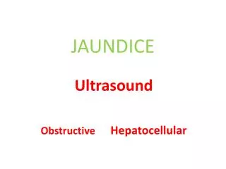
For diagnosis of cause: • USG of whole abdomen with special attention to hepatobiliary system and pancreas (sensitivity 75-90%,specificity 80-100%) • ERCP • MRCP • CT scan • Radioisotope scanning • PTC • Endoscopic ultrasound • Routine investigation: For general anaesthesia: • CBC • Urine R/E • RBS • Serum creatinine • CXR • ECG
For staging • CXR • CT scan • MRI • PET-scan • Bone scan-if symptoms present