Download
1 / 27
370 likes | 961 Vues
FLEXOR TENDON INJURIES James M. Steinberg D.O. Garden City Hospital. Introduction. One of the most common soft tissue injuries of the hand Repair of flexor tendon injuries continues to be a challenging problem Appreciation of flexor tendon anatomy is a must prior to any repair

E N D
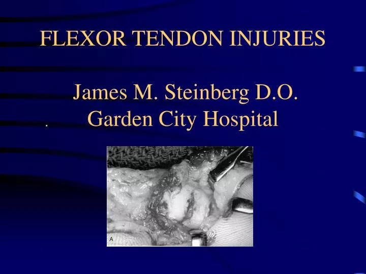
FLEXOR TENDON INJURIES James M. Steinberg D.O.Garden City Hospital
Introduction • One of the most common soft tissue injuries of the hand • Repair of flexor tendon injuries continues to be a challenging problem • Appreciation of flexor tendon anatomy is a must prior to any repair • Repairs used to perform so poorly referred to as surgical no-man’s-land
Tendon Anatomy • Fascicles of long, narrow, spiraling bundles of tenocytes and type I collagen fibers • Fascicles covered by thin visceral and parietal adventitia, paratenon • Paretenon contains fluid similar to synovial fluid • Flexor tendons are enclosed in sheaths lined by visceral and parietal synovial layers • Attached to the sheath weakly by filmy mesenteries composed of vincula
Pulley System • Overly the synovial sheath • Includes: palmar aponeurosis (PA), five annular pulleys (A1-A5), three cruciate pulleys (C1-C3)
Pulley System • PA pulley improves mechanical efficiency of sheath system • Annular pulleys prevent tendon bowstringing • Cruciate pulleys collapse to permit annular pulleys to approximate each other during flexion • Thumb has 2 annular pulleys (A1 at MCP and A2 at IP) and an oblique pulley (lies between A1 and A2) • Oblique pulley is the most important functionally, loss causes decrease in IP motion
Flexor Digitorum Superficialis • Originates from the medial epicondyle, coronoid process, and palmar proximal radius • Superficialis muscle divides into 4 bellies in mid forearm allowing for independent flexion at PIP • Four tendons arise in mid forearm and pass through the carpal tunnel palmar to the profundus tendons • FDS and the intrinsic muscles combine for forceful flexion
Flexor Digitorum Superficialis • At proximal third of prox. phalanx the FDS splits to pass around the profundus (FDP) • Two slips reunite deep to the profundus in a region known as Camper’s Chiasma
Flexor Digitorum Profundus • Originates at proximal 2/3 of the ulna and interosseous membrane • Muscle divides in mid forearm into 2 bellies • radial belly: profundus tendon to index finger • ulnar belly: profundus tendon to long, ring, middle • Tendons pass through the split in the FDS and insert into the base of the prox. 1/3 of the distal phalanges • FDP is the primary digital flexor
Flexors of the Thumb • Flexor pollicis longus (FPL) flexes the IP joint • Flexor pollicis brevis (FPB) flexes the MCP joint • FPL travels within the carpal tunnel
Flexor Zones • Zone I: FDS insertion to FDP insertion • Zone II: A1 pulley to the insertion FDS (No Man’s Land) • Zone III: Distal border of the transverse carpal ligament to A1 pulley • Zone IV: Transverse carpal ligament, (within the carpal Tunnel) • Zone V: Proximal border of the transverse carpal ligament to musculotendinous junctions
Diagnosis • Examiner maintains the other digits in full extension • FDS function: assessed with independent active flexion of the PIP joint • FDP function: determined by active flexion of the DIP joint • FPL function: active flexion of the IP joint of the thumb
Diagnosis • Abnormal resting position of the hand may indicate flexor tendon injury • Squeeze flexor muscles in the forearm • Helpful in the unconscious or noncompliant patient
Tendon Healing • inflammatory phase 48-72hrs • fibroblast/collagen producing phase 5-28 days • remodeling phase which continues for about 112 days
Tendon Healing • inflammatory phase 48-72hrs • fibroblast/collagen producing phase 5-28 days • remodeling phase which continues for about 112 days
Tendon Repair • Primary or delayed primary closure advocated for all zones • Contraindications for primary repair: • contaminated wounds • severe crush or segmental tendon injuries • loss of palmar skin • extensive damage to pulley system
General Considerations of Repair • Knowledge of digit’s position at time of injury • Adequate exposure of proximal and distal tendon ends • mid lateral or palmar zig-zag inscions
Characteristics of an Ideal Repair • Easy placement of suture • Secure knots • Smooth juncture of tendon ends • Minimal gapping at repair site • Minimal interference with tendon vascularity • Sufficient strength throughout healing to allow for early ROM
Repair • Tenorrhaphy in zones I and II most demanding • Numerous techniques have been described • strength of repair is proportional to the number of suture strands that cross repair site • locking loops contribute little strength • repairs rupture at suture knots • synthetic 3-0 or 4-0 braided suture works best
Techniques • Bunnell stitch • Crisscross stitch • Mason-Allen stitch • Becker bevel repair • Kessler grasping stitch • Modified Kessler • Tajima modification of Kessler (double knots)
Zone I Repairs • Vinculum longus is usually intact preventing retraction proximal to the A4 pulley • More than 1cm of profundus advancement results in unacceptable flexion contractures • Need at least 1cm of the distal stump FDP for primary repair--consider insertion into the distal phalanx
Zone II Repairs • Laceration usually between the A2 and A4 pulleys • Often has significant proximal retraction • wrist and MCP at max. flexion • milk flexor muscle bellies • 1 or 2 passes with a tendon retriever under the A2 pulley
Tendon Repair • Studies by Wade and Lin have demonstrated that a running or locked epitendinous sutures increases tensile strength of the repair • Sheath repair remains controversial • repair provides nutrition with synovial fluid • Lister and Tonkin found no benefit with closure • Principles of repair applied to all zones
Partial Tendon Lacerations • Bishop etal. demonstrated that tendon lacerations of 60% or less should not be sutured • Klienert etal. based on cross sectional area: • < 25%: trim edges • 25-50%: repair with simple suture • >50%: repair with modified Kessler
Postoperative Management • Dorsal splint: • 20-30 degrees of palmar flexion at wrist • 45-70 degrees palmar flexion at MCP • extended IP joints • Dynamic splint (Kleinert): • rubber band attached to finger nail • allows for passive flexion against which the patient actively extends
Standard Rehabilitation Protocol • Dorsal splint for 6 weeks • Passive exercises at day 3-4 • Gentle active exercises in 3-4 weeks • Active extension out of splint at 6 weeks • Resistive exercises at 8 weeks
Complications • Rupture • Tendon adhesions • Triggering • Bowstringing
Summary • A thorough understanding of tendon anatomy and physiology, atraumatic surgical technique, and a well designed post-op therapy regiment are a must • Most hand surgeons advocate a four strand core stitch along with a continuous peripheral epitendinous suture • Studies by Gualt, Ikuta, and Savage revealed good to excellent results in 69-90% of patients • Rapid advances continue to occur in flexor tendon surgery, and better techniques will lead to improved outcomes