Download
1 / 31
410 likes | 833 Vues
Fundamentals II: Introduction to Bacteriology and Bacterial Structure. Janet Yother, Ph.D. Department of Microbiology jyother@uab.edu 4-9531. Learning Objectives. Fundamental properties of prokaryotes Basic structures of bacteria Gram-positive vs Gram-negative bacteria.

E N D
Fundamentals II:Introduction to Bacteriology and Bacterial Structure Janet Yother, Ph.D. Department of Microbiology jyother@uab.edu 4-9531
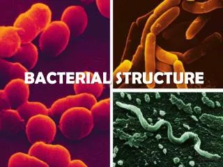
Learning Objectives • Fundamental properties of prokaryotes • Basic structures of bacteria • Gram-positive vs Gram-negative bacteria
Domains (Kingdoms)Based on evolutionary relationships • Eukaryote (Plants, Animals, Protists, Fungi) • Eubacteria (Eubacteria) • Archaea (Archaea)
Bacterial Nomenclature • Kingdom Prokaryotae • Division Gracilicutes • Class Scotobacteria • Subclass • Order Spirochaetales • Family Spirochaetaceae • Tribe • Genus Borrelia • Species Borrelia burgdorferi
BACTERIAL CELL • 50% protein • 20% nucleic acids (10x more RNA than DNA) • 10% polysaccharides • 10% lipids
Bacterial Chromosomes oriC • Single, circular, double-stranded DNA (exception - borrelia = linear) • Replication begins at unique point; bidirectional • Haploid (1 to 4 copies depending on growth rate) • 600 to >5000 kb* in size (smaller = more dependent on host/environment) • Up to 1 mm in length; supercoiled • Contained in nucleoid oriC * ~1 kb/gene
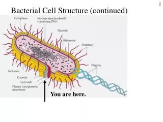
Bacterial Nucleoids • Chromosomal DNA (60%; 2-3% dry wt of cell) + RNA (30%) + Protein (10%) • No nuclear membrane • No histones (~6 chromosome-associated basic proteins involved in determining chromosomal structure) • Polyamines (e.g., spermidine and putrescine) neutralize negative charges on phosphates • Haploid chromosome in cytoplasm • 1 to 4 nuclear bodies/cell, number depends on growth rate (faster = more) • Can be membrane-associated (during cell division) Escherichia coli Electron microscopy Bacillus cereus Light Microscopy 2500x Feulgen strain Jawetz Med Micro 25e
Extrachromosomal DNA • Plasmids - Replicate in cytoplasm, independent of chromosome. • Double-stranded DNA; usually circular (borrelia = linear) • Few to several hundred kb • Few to several hundred copies per cell • Conjugative (F, R), antibiotic resistance, metabolic, virulence • Bacteriophage - virus; • replicates in cytoplasm or integrates into into chromosome • can contribute to virulence
Cytoplasmic Membrane • Lipid bilayer • Permeability barrier • Active transport • Electron transport • Oxidative phosphorylation • Photosynthesis • Affected by antibacterials • Detergents • Polymyxins (damage PE-containing membranes) • Ionophores (disrupt membrane potential)
Cell Wall • Shape • Barrier (osmotic resistance) • Comprised of highly crosslinked peptidoglycan • Affected by antibacterials (e.g, b-lactam antibiotics, lysozyme) • Basis for gram-stain
Peptidoglycan • Backbone of N-acetyl glucosamine and N-acetyl muramic acid • Cross-linked by peptide bridges at MurNAc http://employees.csbsju.edu/hjakubowski/classes/ch331/cho/peptidoglycan.gif
Peptidoglycan http://de.wikipedia.org/wiki/Peptidoglycan
Hydrolases (lysosyme, mutanolysin, e.g.) cleave Transglycosylases (TG) link Amidases (autolysins, e.g.) cleave Transpeptidases (TP) link. Peptidoglycans [GlcNAc-MurNAc]n [GlcNAc-MurNAc]n L-ala D-glu L-lys (gly)n D-ala PG structures vary between/among Gm+ and Gm-. This = Gm+. L-ala D-glu L-lys (gly)n D-ala -lactams resemble TP substrates, block crosslinking of growing chain
[GlcNAc-MurNAc]n R L-ala D-glu L-lys D-ala D-ala NH Transpeptidase CH C O CH2 -lactam ring CH3 HN C O CH3 HC NH HOOC CH C O CH N S C HC (CH3)2 HOOC Benzylpenicillin (penicillin G) Terminal D-ala-D-ala non-crosslinked peptidoglycan -lactams and Peptidoglycan Crosslinking
Gram Stain • Gram’s crystal violet (CV) • Potassium-iodide (KI) • Ethanol - decreases hydration of cell wall • Wash CV-I complexes trapped in thick cell walls (cells remain purple = gram-positive) • Safranin (red) thin cell walls don’t retain CV-I complexes, counterstained with safranin (red = gram-negative)
Exceptions to gram-positive / gram-negative staining • Mycoplasmas - no cell wall. • Mycobacteria - lipid interferes with stain • Detected with acid fast stain (carbol fuschin retained following decolorization with HCl/EtOH) Both are related to gram-positives, based on genetic analyses (rRNA sequence)
Gram-positives • Cytoplasmic Membrane • Cell wall • Lipoteichoic acid • Teichoic acid • Proteins
Gram-positive Cell Walls • Thick peptidoglycan (10 to 100 nm) • Wall teichoic acids (WTA) - repeating units of phosphodiester-linked (negative charge) glycerol or ribitol backbone + side chains (D-ala, glucose). Covalently linked to PG (MurNAc) R1 = H or Ala; R2 = H or Glc
Gram-positive Teichoic Acids • Wall Teichoic Acids (WTA) – covalently linked to PG • Lipoteichoic acids (LTA) – similar to WTA but anchored to cytoplasmic membrane lipids; phosphodiester-linked (negative charge) • LTA and WTA • ion binding • charge maintenance • • membrane integrity • • adherence • • anchor proteins • Cell walls - inflammation
Gram-negatives • Cytoplasmic membrane • Cell Wall • Outer membrane • Lipopolysaccharide • Proteins
Gram-negatives • Cell Wall • Thin peptidoglycan (1 layer; 2 nm) • No WTA or LTA • Periplasmic space - digestive and protective enzymes; transport • Outer membrane (OM) - blocks entry of large molecules (>800 Da). Not typical lipid bilayer. • Attached to PG by lipoprotein • Lipopolysaccharide (LPS) - forms outer leaflet of OM • OM proteins – transport; porins allow passive diffusion of low MW hydrophilic compounds (sugars, amino acids) OmpF
Endotoxin - toxic shock; fever. leukopenia, hypotension, acidosis, DIC, death Lipopolysaccharide (LPS) (OM)-Lipid A --- core polysaccharide --- O Ag polysaccharide varies with strain 3 - 4 sugars/repeat Up to 25 repeats serotyping toxic properties varies with species HM HM MM LM
Gram-negative Surface (Cytoplasmic Membrane)
Optional Features (Gram +/-) • Capsules - polysaccharide or protein (usually covalently linked to peptidoglycan) • Antiphagocytic (block C3b deposition or recognition), attachment • Surface Proteins - anchored in CM, OM, CW • Antiphagocytic, attachment • Flagella - protein. Rotates to propel cell. • Motility, chemotaxis, virulence (H-antigen) Flagella - peitrichous Flagella - EM capsules - colony capsules - microscope Flagella - unipolar
Optional Features (Gram +/-) • Pili - protein. Shorter, narrower than flagella. • Common - peritrichous; attachment • F (sex) - single; gene transfer (conjugation; gram -) • Toxins - excreted; act on host cells; Clostridium botulinum; Vibrio cholerae • Enzymes - hyaluronidase, proteases, DNases • Endospores - dehydrated cells; Clostridium, Bacillus species (gram +) F-pilus