Download
1 / 1
10 likes | 75 Vues
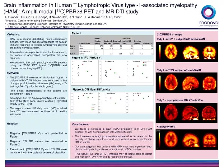
Brain inflammation in Human T Lymphotropic Virus type -1-associated myelopathy (HAM): A multi modal [ 11 C]PBR28 PET and MR DTI study. R Dimber 1 , Q Guo 2 , C Bishop 1 , R Newbould 1 , R N Gunn 1 , E A Rabiner 1,2 , G P Taylor 3 , 1 Imanova, Centre for Imaging Sciences, London UK ,

E N D
Brain inflammation in Human T Lymphotropic Virus type -1-associated myelopathy (HAM): A multi modal [11C]PBR28 PET and MR DTI study R Dimber1, Q Guo2, C Bishop1, R Newbould1, R N Gunn1, E A Rabiner1,2,G P Taylor3, 1Imanova, Centre for Imaging Sciences, London UK, 2 Centre for Neuroimaging Sciences, Institute of Psychiatry, King’s College London,UK, 3 St Mary's Hospital, Imperial College Healthcare NHS Trust, UK Objective: HAM is a chronic debilitating neuro-inflammatory disease, with tissue damage attributed to the cellular immune response to infected lymphocytes entering the central nervous system. Inflammation has a predilection for the thoracic cord, but cases of generalized encephalitis are also reported. We examined the brain pathology in HAM patients using the TSPO PET ligand [11C]PBR28 and diffusion tensor imaging (DTI). Methods: The [11C]PBR28 volumes of distribution (VT) of 3 patients with HTLV1 infection was compared to that of a group of 8 healthy volunteers (HV) using a Z-test (age 56±7 yrs for the whole group) The clinical characteristics of the patients are presented in Table 1 All subjects had the Ala/Ala phenotype of the rs6971 SNP of the TSPO gene, known to affect [11C]PBR28 affinity for the TSPO The regional mean diffusivity index (MD) obtained from DTI was compared to those of 2 healthy volunteers Results: Regional [11C]PBR28 VT s are presented in Figure 1 Regional DTI MD values are presented in Figure 2 Elevations in [11C]PBR28 VT and DTI MD were consistent with the patients degree of disability [11C]PBR28 VT maps Subj 1 - HTLV 1 subject with severe HAM Subj 2 - HTLV1 subject with mild HAM Subj 3 – asymptomatic HTLV1 infection Average of HVs Conclusions: We found a increases in brain TSPO availability in HTLV1 HAM patients, as well as increases in DTI Mean Diffusivity The increases in imaging parameters appeared to be related to the severity of clinical symptoms, and were absent in an asymptomatic HTLV1 carrier Our data suggests that patients with HAM may have significant sub-clinical brain pathology, absent asymptomatic HTLV1 carriers [11C]PBR28 PET and MR DTI imaging may be useful tools to detect and monitor HTLV1 HAM and its response to therapy. Table 1 Figure 1: [11C]PBR28 VT Brain Stem Cerebellum Thalamus Figure 2: DTI Mean Diffusivity Brain Stem Cerebellum Thalamus