1 / 1

Figure e-2

Figure e-2. A. B. C. D. E. F.

dot
Télécharger la présentation

Figure e-2

An Image/Link below is provided (as is) to download presentation Download Policy: Content on the Website is provided to you AS IS for your information and personal use and may not be sold / licensed / shared on other websites without getting consent from its author. Content is provided to you AS IS for your information and personal use only. Download presentation by click this link. While downloading, if for some reason you are not able to download a presentation, the publisher may have deleted the file from their server. During download, if you can't get a presentation, the file might be deleted by the publisher.

E N D

Presentation Transcript


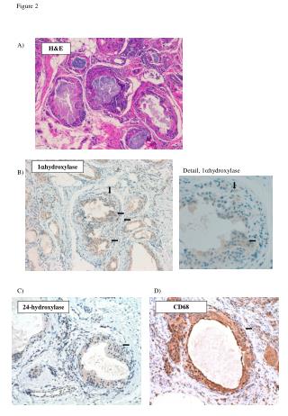
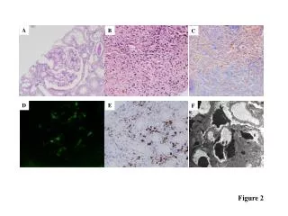
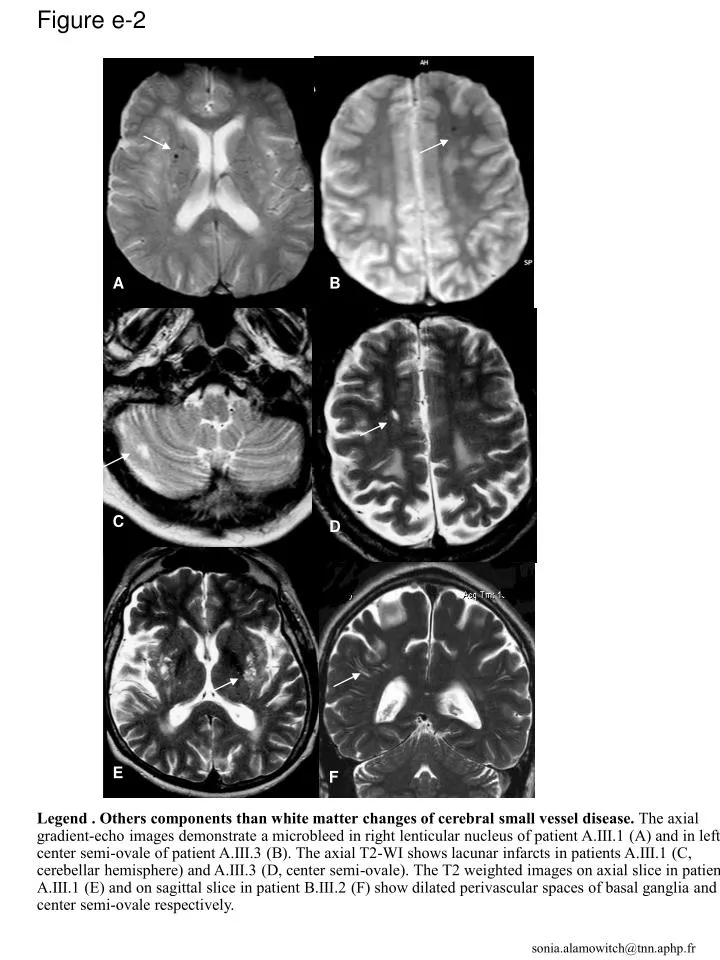
  1. Figure e-2 A B C D E F Legend . Others components than white matter changes of cerebral small vessel disease. The axial gradient-echo images demonstrate a microbleed in right lenticular nucleus of patient A.III.1 (A) and in left center semi-ovale of patient A.III.3 (B). The axial T2-WI shows lacunar infarcts in patients A.III.1 (C, cerebellar hemisphere) and A.III.3 (D, center semi-ovale). The T2 weighted images on axial slice in patient A.III.1 (E) and on sagittal slice in patient B.III.2 (F) show dilated perivascular spaces of basal ganglia and center semi-ovale respectively. sonia.alamowitch@tnn.aphp.fr

More Related