Download
1 / 22
220 likes | 386 Vues
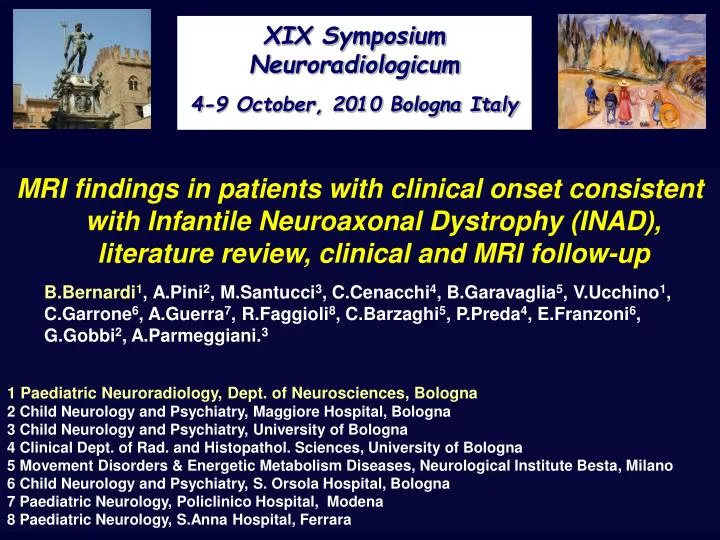
XIX Symposium Neuroradiologicum 4-9 October, 2010 Bologna Italy. MRI findings in patients with clinical onset consistent with Infantile Neuroaxonal Dystrophy (INAD), literature review, clinical and MRI follow-up 1 Paediatric Neuroradiology, Dept. of Neurosciences, Bologna

E N D
XIX Symposium Neuroradiologicum 4-9 October, 2010 Bologna Italy MRI findings in patients with clinical onset consistent with Infantile Neuroaxonal Dystrophy (INAD), literature review, clinical and MRI follow-up 1 Paediatric Neuroradiology, Dept. of Neurosciences, Bologna 2 Child Neurology and Psychiatry, Maggiore Hospital, Bologna 3 Child Neurology and Psychiatry, University of Bologna 4 Clinical Dept. of Rad. and Histopathol. Sciences, University of Bologna 5 Movement Disorders & Energetic Metabolism Diseases, Neurological Institute Besta, Milano 6 Child Neurology and Psychiatry, S. Orsola Hospital, Bologna 7 Paediatric Neurology, Policlinico Hospital, Modena 8 Paediatric Neurology, S.Anna Hospital, Ferrara B.Bernardi1,A.Pini2, M.Santucci3, C.Cenacchi4, B.Garavaglia5, V.Ucchino1, C.Garrone6, A.Guerra7, R.Faggioli8, C.Barzaghi5, P.Preda4, E.Franzoni6, G.Gobbi2, A.Parmeggiani.3
Infantile neuroaxonal dystrophy (INAD) • Rare neurodegenerative disorder involving axons in both central and peripheral nervous systems. • Infantile onset between 6 mos. and 2 yrs. Arrest and rapid regression of motor and cognitive developmental milestones, hypotonia evolving into spastic tetraplegia, cerebellar ataxia and early visual impairment with abnormal visual evoked potentials. • Chronic denervation confirmed by EMG. • Epilepsy has been reported in few cases, EEG can show high-voltage fast rhythms. • Pathological hallmarks: axonal swelling and spheroid bodies (SB) in the pre-synaptic terminals. Nevertheless, SB can be found in other degenerative disorders and are not detected in all tissue specimens of INAD patients.
Infantile neuroaxonal dystrophy (INAD) • 80% of INAD had mutations in the PLA2G6 gene (22q12.3-q13.2) encoding a calcium-independent phospholipase A2 enzyme. • More recently, it has come to light that mutations in the same gene are responsible for some cases of idiopathic neurodegeneration with brain iron accumulation (NBIA). • Evidence that a large percentage of INAD patients have a mutation in the PLA2G6 gene, allows molecular diagnosis and often negates the need for biopsy, an invasive procedure. • On the other hand, the genetic analysis of some patients with typical clinical and pathological features of INAD are negative for PLA2G6 mutations, suggesting the possibility of a genetic heterogeneity for these INAD phenotypes.
PURPOSE • We retrospectively reviewed the MR studies at the onset and during the follow-up of 8 patients in whom clinical and imaging onset met the typical criteria for INAD. The authors compared these data with those reported previously in literature. • Aim of the study was to define if MR features are useful and sufficient in differentiating INAD from other diseases with a similar clinical presentation, guiding clinicians towards appropiate molecular diagnostic testing and to discuss which are the earliest and most important features suggesting INAD diagnosis.
Table 1Clinical features, biopsy and PLA2G6 mutation screening results
INAD: MR findings • MR signs investigated were: • extension & topography of cerebellar atrophy (inclusive criterion) • cerebellar T2 hyperintensity • optic pathway atrophy. • T2 signal of the GP and substantia nigra/subthalamic nuclei • cerebral white matter abnormalities & cerebral atrophy • corpus callosum changes • The MR follow-up of 7 patients included: H+MRS, diffusion MR Imaging (DWI), and diffusion tensor (DTI) analysis.
Cerebellar progressive atrophy and T2 hyperintensity • Diffuse and progressive cerebellar atrophy in all 8 patients. • High T2 signal of the cerebellar cortex in six of eight patients. • Patient 1 with a heterozygous variant of the PLA2G6 gene and + biopsy had a mild cerebellar hyperintensity not progressive and not described in the first of the 3 MRI. • Patient 4 with post mortem pathological INAD diagnosis showed a clearly detectable high T2 of the cerebellar cortex as early as the 1° MRI at 21 months of age and even higher in the follow up.
Patient 1 Female, age of onset 12 months. Biopsy: SB + DNA: heterozygous indeterminate variant of PLA2G6 gene. 3rd MR, 24 months old FLAIR T2WIA FSE T2WI Cr mI Cho NAA MRS Single voxel left hemisphere cerebellar cortex FSE T2WI ADC Patient 1 Cerebellar atrophy, high T2 signal of the cerebellar cortex A FLAIR T2WI B FSE T2WI C FSE T2WI Cr mI MRS Single voxel Left cerebellar hemisphere NAA Cho
T1W Patient 4 T1W T1W • 1° MRI (A,B ) at 21 months of age, • 2° MRI (C,D) at 28 months of age, • 3° MRI (E,F,G) at 5 years and 7 months of age. Progressive cerebellar and corpus callosum atrophy, cerebellar cortex hyperintensity. A C E F B D T2W T2W T2W G PDW T1WI
Cerebellar T2 hyperintensity • High T2 of the cerebellar cortex was also present in patients 2,5,6 and 8 without genetic or biopsy results supporting INAD diagnosis. The first had negative PLA2G6-mutation and clinical and MRI follow-up that became inconsistent with INAD diagnosis. The others were negative for PLA2G6 and PANK2 mutations. • Patient 3 withoutgenetic evaluation for INAD and with negative biopsy as well as patient 7 with negative genetic investigation and biopsy, did not show any significant cerebellar hyperintensity. • T2 cerebellar cortex hyperitensity, initially considered as pathognomonic of the disease, it is not always present, and it may appear only in a later phase, likely resulting from gliosis. • In our group of patients: • Clinical onset from 4 to 15 months of age • Median age of first MRI 17.8 months (range 10 to 23 months)
Patient 81° MRI: 17 monthsMale, age of clinical onset 12 months: arrest acquisition milestones, ataxia, hypotonia, pyramidalism, cataractsNegative PLA2G6 and PANK2; biopsy negative FLAIR T2WI FSE-IR FLAIR T2WI Cerebellar atrophy high T2 signal of the cerebellar cortex
Cr Cho mI Patient 8 NAA MRS LC MODE Exp. DWI
Marinesco–Sjögren : hyperintensity of the cerebellar cortex, widened cerebellar fissures, and an enlarged fourth ventricle in the absence of basal ganglia changes Harting, I. et al. Neurology 2004;63:2448-2449 Cerebellar T2 hyperintensity, even if it is rare, is not a specific feature of INAD but likely a common finding in different degenerative disorders.
Patient 3 Female, age of onset 12 months Biopsy: negative for SBDNA: PLA2G6 not investigated2° MRI at 3 yrs and 3 months: cerebellar atrophy, mild T2 hyperintensity of the cerebellar cortex, optic atrophy. Patient 3. Cerebellar atrophy, mild T2 hyperintensity of cerebellar cortex, optic atrophy
Patient 6 5 year-old Skin biopsy negative DNA not investigated Low GP signal GP GP 3 Tesla MR Unit FSE T2WI 5 year-old age-matched normal controls
Table 2 MR findings +Changes present (+ to ++++ indicating grading); - Changes absent; B: Changes borderline. CH: cerebellar hemisphere; V: vermis; PV: periventricular WM; BG: basal ganglia and internal capsule; Corpus callosum: A: all CC; G genu; B: body; S: splenium corpus callosum; GP: globus pallidus; SN: substantia nigra; STN: subthalamic nuclei.
INAD MR relevance of MR diagnostic criteria • Since absence of either PLA2G6 mutations or pathological evidence may not exclude INAD strongly suggested by clinical features, the evaluation of MR diagnostic criteria has an increased relevance. • In clinical context consistent with the disease, progressive cerebellaratrophy, particularly but not exclusively if associated with early onset of cerebellar cortex high T2, strongly suggests diagnosis of INAD. • Optic atrophy, was evident in patients 1 and 4, both with a definitive diagnosis of INAD but it was also noted in patient 2 and less evident in patients 3 and 7.Although detectable in many other diseases, its presence increases the need to rule out INAD. • GP T2 hypointensity noted only in patient 6, is reported in NBIA patients with PANK2 mutation or in rare atypical NBIA mutations in the PLA2G6 gene. The literature does not consider this sign a crucial feature for early diagnosis of INAD.
INAD MR relevance of MR diagnostic criteria • High ADC in the cerebellar cortex/subcortical WM of all the 7 patients with DWI indicates neuronal loss with increased mobility of water molecules. • All the 7 patients with single voxel 1H-MRS localized in the cerebellum, showed a decreased NAA/creatine ratio. • Patient1, the only having both, a genetically and pathologically confirmed diagnosis of INAD and Spectroscopy, had an associated high mI/Cr ratio in the same cerebellar voxel. • High mI/Cr ratio and absence of lactate also resulted in the cerebellar cortex of 5 the other 6 patients (2,5,6,7,8) without genetic or pathologic evidence of INAD, excluding patient 3 who had a slight lactate peak and a normal mI/Cr ratio.
CONCLUSIONS • Our study confirms that MRI should be considered a standard component of the diagnostic evaluation for INAD and other neurodegenerative disorders. • Cerebellar progressive and diffuse atrophy appears as the most characteristic MR finding in INAD and it was an inclusive criterion in our study. • The limited number of our genetically or pathologically positive patients, does not allow us to confirm that cerebellar cortex T2 hyperintensity is not an obligatory finding of the disease, because it is not always present. • The detection of this sign in patients with negative genetic or pathologic investigations for INAD, confirms that it could be a common MRI feature of many different degenerative conditions.
CONCLUSIONS • The increased diffusion suggests a neuronal loss as well as the low NAA/Cr common finding in the cerebellar H+MRS of all our cases. • Increased mI/Cr suggests astrogliosis a common pathologic feature detectable in late stages of different diseases. • These newer neuroimaging technologies appear to be useful in detecting abnormalities suggesting the nature of the pathological changes characterizing the disorder, but their utility has not yet been established. • At present, it is difficult on the basis of our data to find a definite correlation among clinical and neuroradiological findings with the genetic mutations; at onset and during follow-up it is necessary to study the same variables in a larger samples of patients with INAD and other neurodegenerative diseases.