Download
1 / 3

E N D
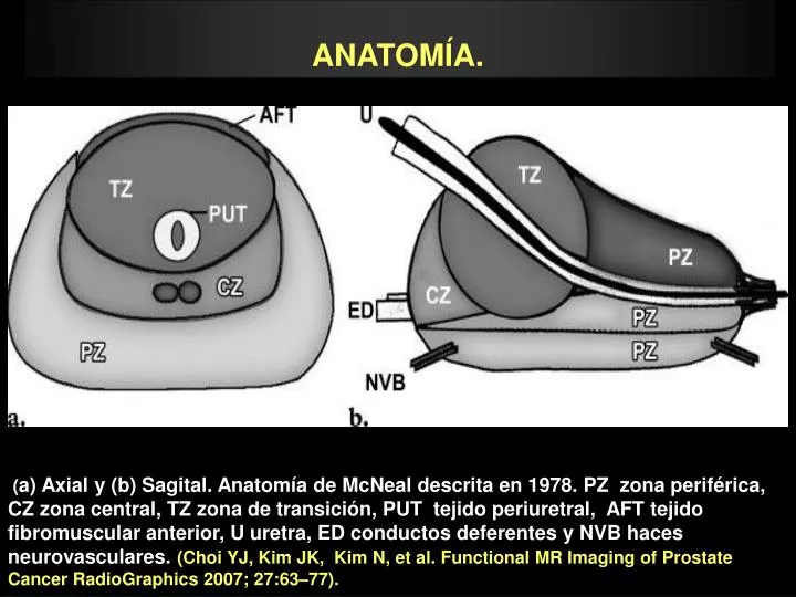
ANATOMÍA. (a) Axial y (b) Sagital. Anatomía de McNeal descrita en 1978. PZ zona periférica, CZ zona central, TZ zona de transición, PUT tejido periuretral, AFT tejido fibromuscular anterior, U uretra, ED conductos deferentes y NVB haces neurovasculares. (Choi YJ, Kim JK, Kim N, et al. Functional MR Imaging of Prostate CancerRadioGraphics 2007; 27:63–77).
ANATOMÍA. A B (A) Axial y (B) Sagital FRSE T2. Zona periférica (flecha roja), zona central (flecha amarilla), zona de transición flecha azul ). Banda estromal fibromuscular (cabeza de flecha roja). Vesícula seminal (cabeza de flecha amarilla).
ANATOMÍA. A B (A) y (B) Coronal FRSE T2. Zona periférica (flecha roja), zona de transición ( flecha azul) y zona periuretral ( cabeza de flecha roja).