Download
1 / 32
480 likes | 1.21k Vues
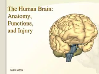
Parts of the Human Brain and their functions. The Human Brain. The human brain is the most complex structure in the known universe. It is the organ that allows you to think, have emotions, move, and even dream – representing the real “you.”

E N D
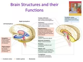
The Human Brain • The human brain is the most complex structure in the known universe. • It is the organ that allows you to think, have emotions, move, and even dream – representing the real “you.” • It is made up of protein, fat, fluid, and weighs approximately 3 pounds. • Traditionally, the brain has been divided into 3 major subdivisions: • The Hindbrain • The Midbrain • The Forebrain
The Hindbrain • The oldest and most primitive level of the brain. • Responsible for our primal instincts and most basic functions. • Damage to this part of the brain will result in death or the need to be sustained on life support.
Brainstem • As the spinal cord enters the brain, it enlarges to form the brainstem. • Attached to the brainstem are the major portions of the hindbrain: the cerebellum, the pons, and the medulla oblangata.
Medulla Oblongata • The medulla is the first structure encountered after leaving the spinal cord. • Plays an important role in vital body functions such as heart rate, respiration and blood pressure which allow you to live. • Functions occur automatically (involuntarily) • A two-way thoroughfare for all sensory and motor nerve tracts between spinal cord and brain • Most of these tracts cross over within the medulla resulting in a crossover of sensory input and motor control
PONS • Meaning ‘bridge’ in Latin, the pons lies above the medulla. • It serves as a bridge carrying nerve impulses between higher and lower levels of the nervous system. • Contains clusters of neurons that help regulate sleep and are involved in dreaming. • Contains motor neurons that control muscles and glands in the face and neck. • Also helps control vital functions, especially respiration.
CEREBELLUm • Meaning ‘little brain’ in Latin, the cerebellum does indeed look like a mini-brain attached to the rear of the brainstem. • The motor coordination centre of the brain – controls muscular movement and coordination. • Specific motor movements are initiated in higher brain centres, but their timing and coordination depend on the cerebellum. • Cats have an especially well-developed cerebellum, helping to account for their graceful and agile movements. • The cerebellum also plays a role in certain types of learning and memory.
Alcohol easily disrupts the coordination of the cerebellum (hence the use of roadside sobriety tests by police). • Physical damage results in severe motor disturbances characterized by jerky, uncoordinated movements, as well as an inability to perform habitual movements such as walking. • Conditions that affect the cerebellum: • Multiple Sclerosis • Stroke • Concussions • Vertigo
The Midbrain • The smallest part of the brain. • Lies just above the hindbrain. • Contains clusters of sensory and motor neurons, as well as many sensory and motor fibre tracts that connect higher and lower portions of the nervous system.
Reticular formation • The brain’s gatekeeper. • Located inside the brainstem, the reticular formation is a finger-shaped structure that extends from the hindbrain up into the lower portions of the forebrain. • The reticular formation acts as a sentry, both alerting higher centres of the brain that messages are coming (the ascending portion) and then either blocking those messages or allowing them to go forward (the descending portion). • Affects consciousness, sleep/wakefulness, and attention.
Some general anaesthetics work by deactivating neurons in the ascending reticular formation, producing a state of unconsciousness in which the sensory impulses that ordinarily would be experienced as pain never register in the sensory areas of the brain involved in pain perception. • In a set of classic experiments in the 1940s, researchers discovered that electrical stimulation of different portions of the reticular formation can produce instant sleep in a wakeful animal and sudden wakefulness in a sleeping animal. • Severe damage to the reticular formation can produce a permanent coma.
The Forebrain • The largest and most recently developed part of the brain. • The most profound biological difference between your brain and that of a lower animal is the size and complexity of your cerebrum. • Consists of 2 large cerebral hemispheres – left and right. • Each cerebral hemisphere is divided into 4 regions, or lobes. • Frontal • Parietal • Temporal • Occipital
Corpus Callosum • Bridge that connects the left and right cerebral hemispheres, passing information from one half of the brain to the other. • Seen as a mass of white matter.
Thalamus • The brain’s sensory switchboard. • Located above the midbrain, the thalamus resembles a pair of egg-shaped structures (one within each cerebral hemisphere). • An important sensory relay station that receives information from all of the senses (except smell) and routes it to higher brain regions that deal with seeing, hearing, tasting and touching. • Also receives some of the higher brain’s replies and directs it to the cerebellum and medulla.
Because of its key role in routing sensory information, disrupted thalamic functioning can produce a highly confusing world for its victims. • Conditions that affect the thalamus: • Schizophrenia
Basal Ganglia • Surrounding and enveloping the thalamus are a group of at least 5 distinct features that are collectively called the basal ganglia. • Whereas the cerebellum is critical for reflexive, automatic, and rapid movements, the basal ganglia plays a critical role in the deliberate and voluntary control of movement. • Conditions that affect the basal ganglia: • Parkinson’s Disease • Tourette’s Disorder • Cerebral Palsy • Attention-Deficit Hyperactivity Disorder (ADHD)
Hypothalamus • Meaning literally “under the thalamus.” • Plays a major role in controlling many different basic biological drives, including sexual behaviour, body temperature regulation, eating/drinking, aggression, and the expression of emotion. • Takes order from other parts of the brain. • Through its connection with the pituitary gland, directly controls many hormonal secretions that regulate sexual development and behaviour, metabolism, and reactions to stress.
Pituitary GLand • Master gland that exerts control over the other glands of the endocrine system. • Tear-shaped structure connected to the hypothalamus. • Releases hormonal secretions to the hypothalamus.
Limbic System • Is made up of a set of structures including the hippocampus, amygdala, and nucleus accumbens. • Helps to coordinate behaviours needed to satisfy motivational and emotional urges that arise in the hypothalamus. • Also plays a role in memory.
Hippocampus • Acts much like a computer processing and storing information. • Involved in forming and retrieving memories. • Processes and stores new and temporary memory information for long-term storage. • Damage can result in severe memory impairment for recent events, and an inability to transfer information from short-term memory to long-term memory. • Conditions that affect the hippocampus: • Amnesia
Amygdala • Means ‘almond’ in Latin (for its shape). • Plays a large role in the production of our emotions, especially aggression and fear. • Its been found to trigger responses to strong emotions such as sweaty palms, chills, increased heart rate and breathing, and stress hormone release.
Nucleus Accumbens • The pleasure centre of the brain. • Rewards and motivation play an important role. • Drugs of abuse, such as cocaine, amphetamines, opiates, nicotine, and alcohol, all stimulate the release of dopamine in the nucleus accumbens, as do naturally occurring rewards such as food and sexual behaviour.
Cerebral Cortex • Outermost layer of the brain (1 cm thick sheet of grey cells); represents 80% of brain tissue. • The crowning achievement of brain evolution as no fish or amphibian has one. • Progression from more primitive to more advanced mammals is marked by a dramatic increase in the proportion of cortical tissue. • the cortex makes up 80% of the human brain. • The CEO of the brain. • Not essential for physical survival, but is essential for quality of living. • Deep fissures (folds) within it are what help divide the brain into left and right hemispheres, and lobes.
Association Cortex • Found within all lobes of the brain. • Referred to as “silent areas” because electrically stimulating them does not give rise to either sensory experiences or motor responses. • Critically involved in the highest level of mental functions including perception, language, and thought.
Frontal Lobe • Located behind your eyes and forehead. • Responsible for speech, emotion, behaviour, movement, decision-making, problem-solving, and planning. • MOTOR CORTEX • Located at the rear of the frontal lobe • Out-going messages • Controls over 600 muscles involved in voluntary body movements • Each cerebral hemisphere governs movement on the opposite side of the body • Specific body areas are represented in different parts of the motor cortex, and the amount of cortex devoted to each area depends on the complexity of the movements that are carried out by the body part
Parietal Lobe • Located at the top and to the rear of your head. • Concerned with the reception and processing of sensory information from the body. • Allows us to process pain, pressure, and other physical sensations and identify objects. • SOMATIC SENSORY CORTEX • Located at the front of the parietal lobe • Incoming messages • Receives sensory input that gives rise to our sensations of heat, cold, touch, and our senses of balance and body movement • As in the case of the adjacent motor cortex, each side of the body sends sensory input to the opposite cerebral hemisphere • Likewise, the amount of cortex devoted to each body areas is directly proportional to that region’s sensory sensitivity
Temporal Lobe • Located just above the ears. • Controls memory, personality, hearing, and language. • WERNICKE’S AREA • Located in the temporal lobe • Named for Carl Wernicke who discovered that damage to this cortical region left patients unable to understand written or spoken speech • Involved in language and comprehension • BROCA’S AREA • Located in the frontal lobe • Named for Paul Broca who discovered that damage to this cortical region left patients with the ability to comprehend speech but not to express themselves in words or sentences • Necessary for normal speech production
Occipital Lobe • Located at the back of your head. • Processes visual stimuli and allows the brain to process light and objects.