Download
1 / 106
1.1k likes | 1.34k Vues
Parts of the brain. Sanjaya Adikari Department of Anatomy. Spinal cord. Foramen magnum. Central Nervous System (CNS). Skull. Vertebral column. Meninges. Dura mater. Arachnoid mater. Pia mater. Dura mater. Arachnoid mater. Ventricle. Pia mater. Ependymal cell layer. 1. 1. 2. 3.

E N D
Parts of the brain Sanjaya Adikari Department of Anatomy
Spinal cord Foramen magnum Central Nervous System (CNS)
Skull Vertebral column
Meninges Dura mater Arachnoid mater Pia mater
Dura mater Arachnoid mater Ventricle Pia mater Ependymal cell layer
1 1 2 3 3 4 4 6 6 5 5 Main divisions of the brain = forebrain 2 + = midbrain = hindbrain
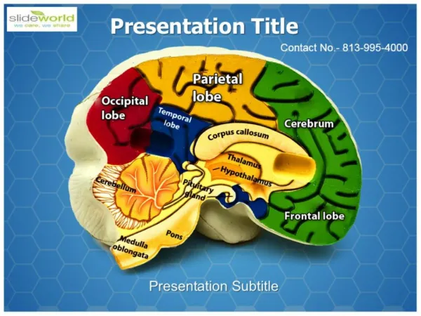
1 3 4 6 5 2 + Cerebrum Diencephalon midbrain = brainstem pons medulla Cerebellum
Cerebrum Cerebrum is the largest part of the brain. It is situated in the anterior and middle cranial fossae and the whole concavity of the vault of the skull Has two parts; • Cerebral hemispheres • Left & right cerebral hemispheres • Diencephalon • Consists of thalamus, hypothalamus etc.
Diencephalon thalamus hypothalamus Spinal cord Mid-sagittal section of brain
Diencephalon thalamus hypothalamus Spinal cord
Cerebral hemispheres • Largest part of the brain • Separated by a deep mid-sagittal fisure called longitudinal cerebral fissure • The fissure contains the falx cerebri and anterior cerebral arteries • Tentorium cerebelli separates cerebral hemispheres from the cerebellum
Falx cerebri Anterior cerebral arteries Dura mater Tentorium cerebelli Corpus callosum
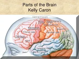
Sulci and Gyri • To increase the surface area of the brain the cerebral hemispheres are thrown into folds - gyri • The gyri are separated from each other byfissures - sulci • Hemispheres are divided into lobes (named according to the cranial bones under which they lie) by main sulci • Central • Parieto-occipital • Lateral
Central sulcus Parietal Lobe Frontal Lobe Parieto-occipital sulcus Lateral sulcus OccipitalLobe Temporal Lobe
Main gyri • Precentral gyrus • Postcentral gyrus • Superior/middle/inferior frontal gyri • Superior/middle/inferior temporal gyri • Cingulate gyrus • Parahippocampal gyrus
Precentral gyrus Postcentral gyrus Superior, middle, inferior frontal gyri Superior, middle, inferior temporal gyri
Cingulate gyrus Parahippocampal gyrus
Homework Draw a labelled line diagram to illustrate a mid-sagittal section of the brain including the brain stem. Draw a labelled line diagram to illustrate a horizontal section of the cerebrum through the head of the caudate nucleus. Study the above diagrams before you come for the next lecture on parts of the brain
Gray matter and white matter Gray matter White matter • Gray matter consists of nerve cells • White matter consists of nerve fibres
Gray matter of the cerebral cortex Five types of cells are organized into six cortical layers
Molecular layer External granular layer External pyramidal layer Internal granular layer Ganglionic layer (Internal pyramidal layer) Multiform layer
White matter • Composed of myelinated nerve fibres • Supported by neuroglia • Classified into three groups according to their connections • Commissural fibers • Association fibers • Projection fibers
Commissural fibers Connects corresponding regions of the two hemispheres Corpus callosum, fornix, anterior and posterior commissures Corpus callosum, the largest commissure of the brain, is divided into rostrum, genu, body and the splenium
Body Genu Splenium Rostrum Corpus callosum
Association fibers • Connects various cortical regions within the same hemispheres • Divided into short and long groups • Short association fibers lie immediately beneath the cortex and connect adjacent gyri • Long association fibers are arranged into named bundles - fasciculi
Projection fibers • Afferent and efferent nerve fibers passing to and from the brain stem to the cerebral cortex • Internal capsule, corona radiata, optic radiation
Corona radiata Internal capsule Optic radiation Optic tract Cerebral peduncle Pyramidal tract Internal capsule and corona radiata
Internal capsule Putamen Cerebral peduncle Amygdala
Basal ganglia • Basal ganglia are collection of masses of gray matter within the white matter of cerebral hemispheres Gray matter of cerebral cortex White matter of cerebrum Basal ganglia
Basal ganglia…..cont. • Corpus striatum • Divided into two by internal capsule of white matter • Caudate nucleus • Lentiform nucleus (putamen & globus pallidus) • Amygdaloid • Claustrum
Claustrum External capsule Anterior horn of lateral ventricle Thalamus Head of caudate nucleus Lentiform nucleus (putamen) Internal capsule Lentiform nucleus (globus pallidus) Tail of caudate nucleus
Fiber tracts in the internal capsule Frontopontine Corticobulbar Corticospinal Thalamocortical Parieto/temporo/occipito pontine Visual & auditory
Basal ganglia…..cont. Some definitions include the following also under basal ganglia Subthalamic nucleus Substantia nigra Midbrain
Diencephalon Consists of the following Thalamus Subthalamus Hypothalamus Epithalamus Habenular nucleus Pineal gland
Corpus callosum Fornix Thalamus Mamillary body
Thalamus Epithalamus Subthalamus Hypothalamus
Fornix Roof of 3rd ventricle Interthalamic connection Lentiform nucleus Thalamus Internal capsule Hypothalamus Optic chiasma Pituitary Mammillary body
Thalamus • Large ovoid mass of gray matter • Forms large part of diencephalon • Very important cell station • Receives main sensory tracts (except olfactory pathway) • Integrates information it receives and relays to the cerebral cortex and subcortical regions • Integrates visceral and somatic functions
Hypothalamus • Part of the diencephalon that extends from the optic chiasma to the posterior border of the mammillary bodies • Almost all physiological activities of the body are influenced by hypothalamus • Integration of autonomic functions • Regulation of endocrine functions • Maintaining body homiostasis • Regulation of body temperature and body fluids • Sexual behaviour, emosions, drive to eat and drink
Hypothalamus…. • Contains some important cell groups • Supraoptic nucleus • Paraventricular nucleus • These have axons running down into the posterior lobe of the pituitary gland • Other cell groups deliver their neurosecretions into the hypothalamo-hypophyseal poryal system leading to the anterior lobe of the pituitary gland