Download
1 / 42
540 likes | 857 Vues
Parts of the Brain. Dr Ajith Sominanda Department of Anatomy. Nervous System. Environmental stimuli. Information Processing. Effects. Nervous system and Brain Few facts from your A/Levels or high school biology. Nervous system consists of nerve tissues: neurons and glia

E N D
Parts of the Brain Dr Ajith Sominanda Department of Anatomy
Nervous System Environmental stimuli Information Processing Effects
Nervous system and BrainFew facts from your A/Levels or high school biology • Nervous system consists of nerve tissues: neurons and glia • Brain and spinal cord belongs to CNS • In CNS, macroscopically white and gray matter are identifiable • These white and gray matter are arranged into different areas of the brain and spinal cord
Nervous system and BrainFew facts from your A/Levels or high school biology • In a Fresh brain or Spinal cord., • White is due to myelinated (protein +l ipid); nerve fibers or Axons • Gay is due to cells; neurons & glia But in imaging techniques gray and white may look different
Nervous system and BrainFew facts from your A/Levels or high school biology
Brain in the cranial cavity • video
Terms in Neuroanatomy encountered in your A/Ls Nervous System • CNS (Brain & Spinal cord) • PNS (Cranial nerves, Spinal nerves, peripheral ganglia) Nerve tissue Neuron Glial cell Neurits (Axons & Dendrits) Synapse Nerve fiber, ganglia & peripheral nerve Gray matter & White matter Development Neuroectoderm, neural tube, neural crest cells Cerebrum Cerebellum Brain Stem (mid brain, pons, medulla) Meningies (Dura mater, arachnoid mater & pia mater) Ventricles CSF
Development of the brain Is the key to understand its structure
Neural Tube Neural crests
Development - Major points • Nervous system develops from an area of ectoderm called neuroectoderm or neural plate of the embryo • Neuroectoderm give rise to neural tube and the peripheral nerves develop from neural crests
Naming different areas of the developing neural tube • Forebrain • Midbrain • Hindbrain
neural tube bends and grow Cephalic flexure Cervical flexure Pontine flexure Embryo Swollen & Folded neural tube
Areas of developing neural tube and their future components Hindbrain Midbrain Forebrain
Thus, brain is the modified cephalic (front) part of neural tube by growth (swelling) and folding
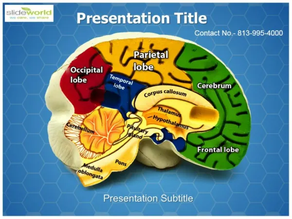
Main parts of the adult brainRevision Forebrain Cerebral hemispheres or cerebrum Diencephalon Thalamus, Epithalamus, Hypothalamus & Subthalamus Midbrain Hindbrain Pons and Cerebellum Medulla
ForebrainCerebrum or Cerebral hemispheres • Represent the largest part of the brain • Has extensively convoluted cerebral cortex • Has internal white matter fiber bundles • Has internal masses of gray matter • Also contains cavities; lateral ventricles • Two hemispheres are connected by a bundle of white matter called corpus callosum
Cerebral Cortex (gray matter) • Contains cells (neurons & glia) • Extensive folding forms sulci and gyri • Large sulci / fissures divides cerebral hemispheres into different lobes
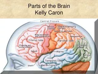
Topography of cerebral hemispheres Supero-lateral surface of the brain • Sulci- Lateral, Central, parieto-occipital • Frontal lobe, Parietal lobe, Occipital lobe & Temporal lobe
Main Sulci and Gyri Draw
Topography of cerebral hemispheres medialsurface of the brain • Cingulategyrus, cuneus, lingulargyrus • Cingulatesulcus, calcarinesulcus
Cerebral White matter • Consists of Axons that connect different parts of the nervous system • These axons are arranged in bundles which can be displayed by dissection
Cerebral White matter Three types of axon bundles (fasciculi) are present in cerebral white matter: • Association fibers Confined to a hemisphere and connects cortical areas within the hemisphere • Transverse or Commissural fibers • Connects 2 hemispheres • Axons runs in corpus callosum and anterior commissure, • Projection fibers Connect cerebral cortex with subcortical structures, brain stem and spinal cord
Cerebral White matter Corpus Callosum (Transverse or Commissural fibers)
Cerebral White matter Corpus Callosum
Cerebral White matter Corona radiata & Internal capsule (Contains projection fibers)
Cerebral White matter More details during the practical sessions
Other internal structures of cerebral hemispheres • Diencephalic structures (Thalamus, hypothalamus, epi thalamus and sub thalamus)
Other internal structures of cerebral hemispheres • Telencephalic gray matter • Corpus striatum (telencephalic gray matter associated with lateral ventricles) • Striatum (caudate nucleus, nucleus accumbens & putamen) • Pallidium (globuspallidus)
Histology of cerebral cortex • Cerebral gray and white matter is (histologically) arranged in layers • Three Histologically different areas can be identified: • Paleocortex (olfactory system) • Archicortex (hipocampal formation) • Neocortex (rest of cerebral cortex) - 3 layers 6 layers
Histology of cerebral cortex Neocortical Histology
Histology of cerebral cortex There are two types of neurons : • Principal neurons • Typical principal cells are pyramidal cells • Atypical principal cells are fusifom cells • Inter neurons
Histology of cerebral cortex Principal neurons connect with other neurons in CNS in 3 ways • Projection neurons/fibers (subcortical areas such as thalamus, corpus striatum, brain stem & spinal cord) • Association neurons/fibers (connects cortical neurons in same hemisphere) • Comissural neurons/fibers (connects cortical neurons in opposite hemisphere )