Download
1 / 59
640 likes | 987 Vues
6 - Lecture Osseous Tissue and Bone Structure. An Introduction to the Skeletal System. Learning Outcomes 6-1 Describe the primary functions of the skeletal system. 6-5 Compare the mechanisms of endochondral ossification and intramembranous ossification.

E N D
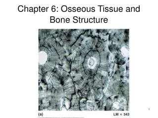
6 - Lecture Osseous Tissue and Bone Structure
An Introduction to the Skeletal System • Learning Outcomes • 6-1 Describe the primary functions of the skeletal system. • 6-5 Compare the mechanisms of endochondral ossification and intramembranous ossification. • 6-6 Describe the remodeling and homeostatic mechanisms of the skeletal system. • 6-7 Discuss the effects of exercise, hormones, and nutrition on bone development and on the skeletal system.
An Introduction to the Skeletal System • Learning Outcomes • 6-8 Explain the role of calcium as it relates to the skeletal system. • 6-9 Describe the types of fractures, and explain how fractures heal. • 6-10 Summarize the effects of the aging process on the skeletal system.
An Introduction to the Skeletal System • The Skeletal System • Includes: • Bones of the skeleton • Cartilages, ligaments, and connective tissues
6-1 Functions of the Skeletal System • Five Primary Functions of the Skeletal System • Support • Storage of Minerals (calcium) and Lipids (yellow marrow) • Blood Cell Production (red marrow) • Protection • Leverage (force of motion)
6-5 Bone Formation and Growth • Bone Development • Human bones grow until about age 25 • Osteogenesis • Bone formation • Ossification • The process of replacing other tissues with bone
6-5 Bone Formation and Growth • Bone Development • Calcification • The process of depositing calcium salts • Occurs during bone ossification and in other tissues • Ossification • Two main forms of ossification • Endochondral ossification • Intramembranous ossification
6-5 Bone Formation and Growth • Endochondral Ossification • Ossifies bones that originate as hyaline cartilage • Most bones originate as hyaline cartilage • There are six main steps in endochondral ossification
Figure 6-10 Endochondral Ossification Enlargingchondrocytes withincalcifying matrix Hyaline cartilage
Figure 6-10 Endochondral Ossification Epiphysis Diaphysis Bone formation
Figure 6-10 Endochondral Ossification Medullary cavity Blood vessel Primary ossification center Superficial bone Spongy bone
Figure 6-10 Endochondral Ossification Medullary cavity Metaphysis
Figure 6-10 Endochondral Ossification Hyaline cartilage Epiphysis Metaphysis Periosteum Compact bone Secondary ossification center
Figure 6-10 Endochondral Ossification Articular cartilage Spongy bone Epiphyseal cartilage Diaphysis
Figure 6-10 Endochondral Ossification Cartilage cells undergoing division and secreting additional cartilage matrix Epiphyseal cartilage matrix LM 250 Osteoid Osteoblasts Medullary cavity
6-5 Bone Formation and Growth • Appositional Growth • Compact bone thickens and strengthens long bone with layers of circumferential lamellae ANIMATION Endochondral Ossification
6-5 Bone Formation and Growth • Epiphyseal Lines • When long bone stops growing, after puberty: • Epiphyseal cartilage disappears • Is visible on X-rays as an epiphyseal line • Mature Bones • As long bone matures: • Osteoclasts enlarge medullary (marrow) cavity • Osteons form around blood vessels in compact bone
Figure 6-11a Bone Growth at an Epiphyseal Cartilage An x-ray of growing epiphyseal cartilages (arrows)
Figure 6-11b Bone Growth at an Epiphyseal Cartilage Epiphyseal lines in an adult (arrows)
6-5 Bone Formation and Growth • Intramembranous Ossification • Also called dermal ossification • Because it occurs in the dermis • Produces dermal bones such as mandible (lower jaw) and clavicle (collarbone) • There are three main steps in intramembranous ossification
Figure 6-12 Intramembranous Ossification Mesenchymal cells aggregate, differentiate into osteoblasts, and begin the ossification process. The bone expands as a series of spicules that spread into surrounding tissues. Osteocyte in lacuna Bone matrix Osteoblast Osteoid Embryonic connective tissue Blood vessel Mesenchymal cell LM 22 Blood vessel Osteoblasts Spicules
Figure 6-12 Intramembranous Ossification Over time, the bone assumes the structure of spongy bone. Areas of spongy bone may later be removed, creating medullary cavities. Through remodeling, spongy bone formed in this way can be converted to compact bone. Blood vessel
6-5 Bone Formation and Growth • Blood Supply of Mature Bones • Nutrient Artery and Vein • A single pair of large blood vessels • Enter the diaphysis through the nutrient foramen • Femur has more than one pair • Metaphyseal Vessels • Supply the epiphyseal cartilage • Where bone growth occurs • Periosteal Vessels • Blood to superficial osteons • Secondary ossification centers
Figure 6-13 The Blood Supply to a Mature Bone Articular cartilage Epiphyseal artery and vein Branches of nutrient artery and vein Metaphyseal artery and vein Periosteum Periosteum Compact bone Periosteal arteries and veins Connections to superficial osteons Medullary cavity Nutrient artery and vein Nutrient foramen Metaphysis Metaphyseal artery and vein Epiphyseal line
6-5 Bone Formation and Growth • Lymph and Nerves • The periosteum also contains: • Networks of lymphatic vessels • Sensory nerves
Figure 6-9 Heterotopic Bone Formation The skeleton of a man with advanced FOP An adult male with FOP, posterior view
6-6 Bone Remodeling • Process of Remodeling • The adult skeleton: • Maintains itself • Replaces mineral reserves • Recycles and renews bone matrix • Involves osteocytes, osteoblasts, and osteoclasts
6-6 Bone Remodeling • Process ofRemodeling • Bone continually remodels, recycles, and replaces • Turnover rate varies: • If deposition is greater than removal, bones get stronger • If removal is faster than replacement, bones get weaker
6-7 Exercise, Hormones, and Nutrition • Effects of Exercise on Bone • Mineral recycling allows bones to adapt to stress • Heavily stressed bones become thicker and stronger • Bone Degeneration • Bone degenerates quickly • Up to one third of bone mass can be lost in a few weeks of inactivity
6-7 Exercise, Hormones, and Nutrition • Normal Bone Growth and Maintenance Depend on Nutritional and Hormonal Factors • A dietary source of calcium and phosphate salts • Plus small amounts of magnesium, fluoride, iron, and manganese
6-7 Exercise, Hormones, and Nutrition • Normal Bone Growth and Maintenance Depend on Nutritional and Hormonal Factors • The hormone calcitriol • Made in the kidneys • Helps absorb calcium and phosphorus from digestive tract • Synthesis requires vitamin D3 (cholecalciferol)
6-7 Exercise, Hormones, and Nutrition • Normal Bone Growth and Maintenance Depend on Nutritional and Hormonal Factors • Vitamin C is required for collagen synthesis, and stimulation of osteoblast differentiation • Vitamin A stimulates osteoblast activity • Vitamins K and B12 help synthesize bone proteins
6-7 Exercise, Hormones, and Nutrition • Normal Bone Growth and Maintenance Depend on Nutritional and Hormonal Factors • Growth hormone and thyroxine stimulate bone growth • Estrogens and androgens stimulate osteoblasts • Calcitonin and parathyroid hormone regulate calcium and phosphate levels
Figure 6-14 Examples of Abnormal Bone Development Pituitary dwarfism Marfan’s syndrome
6-8 Calcium Homeostasis • The Skeleton as a Calcium Reserve • Bones store calcium and other minerals • Calcium is the most abundant mineral in the body • Calcium ions are vital to: • Membranes • Neurons • Muscle cells, especially heart cells
Figure 6-15 A Chemical Analysis of Bone Bone Contains … Composition of Bone Calcium 39% 99% of the body’s Calcium Potassium 0.2% 4% of the body’s Potassium 35% of the body’s Sodium Sodium 0.7% 50% of the body’s Magnesium Magnesium 0.5% Organic compounds (mostly collagen) 33% 80% of the body’s Carbonate Carbonate 9.8% Phosphate 17% 99% of the body’s Phosphate Total inorganic components 67%
6-8 Calcium Homeostasis • Calcium Regulation • Calcium ions in body fluids • Must be closely regulated • Homeostasis is maintained • By calcitonin and parathyroid hormone (PTH) • Which control storage, absorption, and excretion
6-8 Calcium Homeostasis • Calcitonin and Parathyroid Hormone Control • Affect: • Bones • Where calcium is stored • Digestive tract • Where calcium is absorbed • Kidneys • Where calcium is excreted
6-8 Calcium Homeostasis • Parathyroid Hormone (PTH) • Produced by parathyroid glands in neck • Increases calcium ion levels by: • Stimulating osteoclasts • Increasing intestinal absorption of calcium • Decreasing calcium excretion at kidneys • Calcitonin • Secreted by C cells (parafollicular cells) in thyroid • Decreases calcium ion levels by: • Inhibiting osteoclast activity • Increasing calcium excretion at kidneys
Figure 6-16a Factors That Alter the Concentration of Calcium Ions in Body Fluids Factors That Increase Blood Calcium Levels These responses are triggered when plasma calcium ion concentrations fall below 8.5 mg/dL. Low Calcium Ion Levels in Plasma (below 8.5 mg/dL) Parathyroid Gland Response Low calcium plasma levels cause the parathyroid glands to secrete parathyroid hormone (PTH). PTH Bone Response Intestinal Response Kidney Response Kidneys retain calcium ions Rate of intestinal absorption increases Osteoclasts stimulated to release stored calcium ions from bone more Osteoclast Bone calcitriol Calcium absorbed quickly Calcium conserved Calcium released Decreased calcium loss in urine ↑Ca2+ levels in bloodstream
Figure 6-16b Factors That Alter the Concentration of Calcium Ions in Body Fluids Factors That Decrease Blood Calcium Levels HIgh Calcium Ion Levels in Plasma (above 11 mg/dL) These responses are triggered when plasma calcium ion concentrations rise above 11 mg/dL. Thyroid Gland Response Parafollicular cells (C cells) in the thryoid gland secrete calcitonin. Calcitonin Intestinal Response Bone Response Kidney Response Kidneys allow calcium loss Rate of intestinal absorption decreases Osteoclasts inhibited while osteoblasts continue to lock calcium ions in bone matrix less Bone calcitriol Calcium excreted Calcium absorbed slowly Calcium stored Increased calcium loss in urine ↓Ca2+ levels in bloodstream
6-9 Fractures • Fractures • Cracks or breaks in bones • Caused by physical stress • Fractures are repaired in four steps • Bleeding • Cells of the endosteum and periosteum • Osteoblasts • Osteoblasts and osteocytes remodel the fracture for up to a year
6-9 Fractures • Bleeding • Produces a clot (fracture hematoma) • Establishes a fibrous network • Bone cells in the area die • Cells of the endosteum and periosteum • Divide and migrate into fracture zone • Calluses stabilize the break • External callus of cartilage and bone surrounds break • Internal callus develops in medullary cavity
Figure 6-17 Types of Fractures and Steps in Repair REPAIR OF A FRACTURE Fracture hematoma Periosteum Bone fragments Spongy bone of external callus Dead bone Immediately after the fracture, extensive bleeding occurs. Over a period of several hours, a large blood clot, or fracture hematoma, develops. An internal callus forms as a network of spongy bone unites the inner edges, and an external callus of cartilage and bone stabilizes the outer edges.
6-9 Fractures • Osteoblasts • Replace central cartilage of external callus • With spongy bone • Osteoblasts and osteocytes remodel the fracture for up to a year • Reducing bone calluses
Figure 6-17 Types of Fractures and Steps in Repair External callus External callus Internal callus A swelling initially marks the location of the fracture. Over time, this region will be remodeled, and little evidence of the fracture will remain. The cartilage of the external callus has been replaced by bone, and struts of spongy bone now united the broken ends. Fragments of dead bone and the areas of bone closest to the break have been removed and replaced.
6-9 Fractures • Major Types of Fractures • Transverse fractures • Displaced fractures • Compression fractures • Spiral fractures • Epiphyseal fractures • Comminuted fractures • Greenstick fracture • Colles fracture • Pott’s fracture