Download
1 / 71
780 likes | 1.22k Vues
SPLEEN. Mahdi Alemrajabi MD. Faculty member of Iran university of medical sciences Firoozghar general hospital www.alemrajabi.ir. He is a splenetic man.

E N D
SPLEEN Mahdi Alemrajabi MD. Faculty member of Iran university of medical sciences Firoozghar general hospital www.alemrajabi.ir
He is a splenetic man.
In Englysche it is named a mans Splene. A spongious substance lienge under the ribbes on the left side, and it doth make a man to bee mery and to laughe, although melancholy resteth in the splene if there be impediments in it. If any man be splenitike let him use mery company, be joconde, and not to study upon any supernatural thynges.’ (Andrew Boorde; The Breviarie of Helthe, 1547).
Anecdotal reports of splenic surgery began to emerge in the sixteenth century. By the end of the eighteenth century, the vast majority of splenectomies performed were partial, with the majority of patients requiring surgical attention for left upper quadrant stab wounds resulting in partial or total splenic prolapse. By 1877 only 50 splenectomies had ever been performed, with a mortality rate of >70%. Yet by 1900, large series of splenectomies were reported with an operative mortality rate that had already dropped to <40%, and a 1920 report1 of the Mayo Clinic experience with splenectomy noted an 11% mortality rate. Somewhat less dramatic progress has since occurred, with the largest series of laparoscopic splenectomies reporting 1% overall mortality.
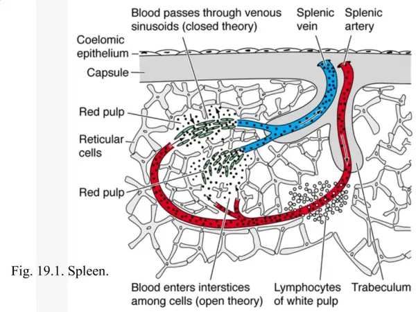
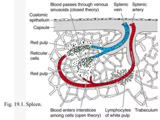
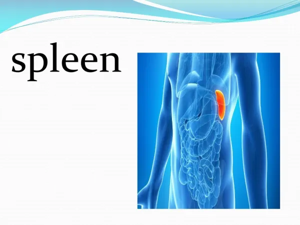
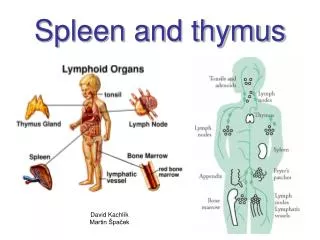
Position of spleen • The spleen lies in the left hypochondrium between the stomach and diaphragm. • It has two ends, three borders and two surfaces.
biggest reticoloendotelial organ accessory spleens have been reported in 20% to 30%, ususally seen at the hilum, greater omentum, and ligaments of the spleen
Beneath ribs 9-11 • Length 7-11 cm • Weight 150 gr • SI 120-480 ml
Causes of splenomegaly • Infection • Bacterial: Typhoid fever, endocarditis, septicemia, abscess • Viral:E-B virus, CMV, and others • Protozoal: Malaria, toxoplasmosis • Hematologic processes • Hemolytic anemia: Congenital, acquired • Extramedullary hematopoiesis: thalassemia, osteopetrosis, myelofibrosis • Neoplasms • Malignant: Leukemia, lymphoma, histiocytoses, metastatic tumors • Benign: Hemagioma, hamartoma • Metabolic diseases • Lipidosis: Niemann-Pick, Gaucher disease • Mucopolysaccharidosis infiltration: Histiocytosis • Congestion • Cirrhosis • Cysts • Miscellaneous
II. Physiologic Functions A. Filtering – splenic blood flow – 300 ml/min 1. removal of abnormal red blood cells – approximately 20 ml of aged RBC are removed daily 2.removal of abnormal WBC, and plateletes
B. Immunologic Function 1. opsonin, tuftesin and properdinproduction 2. antibody synthesis (IgM) 3. protection from infection C. Storage Function 1. plateletes – 1/3 are stored in the spleen 2. in splenomegaly, up to 80% of the plateletesmay be stored in the spleen thrombocytopenia D.Hematopioesis
Hypersplenism • presence of one or more cytopenias in the context of a normally functioning bone marrow. 1-increased destruction of abnormal blood cells occurs in an intrinsically normal spleen (e.g., hemolytic anemias) 2- primary disorders of the spleen resulting in increased sequestration and destruction of normal blood cells (e.g., infiltrative disorders).
Blood cells half life: • RBC 120 days • PMN 6 hrs • Plt 10 days
splenosis • Is the auto transplantation of splenic tissue after splenic trauma • They vary from a few millimeters to several centimeters in diameter • May occur anywhere in the peritoneal cavity • Seldom causes symptoms and is usually discovered as an incidental finding at reoperation • Post splenectomy sepsis has renewed interest in splenosis
INDICATIONS OF SPLENECTOMY More prevalent Urgent trauma Elective ITP
Splenic Trauma: • Most common indication for laparotomy after blunt trauma • Most commonly injured abdominal organ in blunt trauma • Mechanism: • MVC, MCC, falls, PVA, bicycle crashes, sports • Injuries : rapid deceleration-> avulsion along ligaments Efficient energy transfer form chest wall Direct punctures from rib fracture
Diagnosis of Splenic Trauma: • Historically PE: • peritoneal signs (42-72% accurate) • Bruising over LUQ • Kehr sign: left upper quadrant pain, with referred left shoulder pain • Hypotension, tachycardia-> suspicious for hemorrhage, not attributed to other source • Confounding factors: head, spinal cord injury, substance abuse • West et all: development of trauma systems: mortality from delayed/missed recognition of splenic hemorrhage still major cause of preventable death
Diagnosis of Splenic Trauma cont: 2. DPL: • Introduced in 1965 by Root • standard of care for blunt abdominal trauma for 20 yrs • Originally: 10ml blood aspirated=> + • Now: 1L crystalloid infusion=> >100,000 RBCs, 500 WBCs • Sensitivity: 99%, Specificity: 95-98% • Drawback: “nontherapeutic laparotomies”
Diagnosis of Splenic Trauma cont: 3. Ultrasound • Introduced in 1990s • FAST( focused abdominal sonogram for trauma): • noninvasive, rapid, low cost • Presence of intraperitoneal fluid, replaced DPL • OR without CT scan in unstable patient • stable patient: screening for CT scan • Limited by obesity, bowel gas and subcutaneous emphysema - Sensitivity: 90-93%, Specificity: 99%
Diagnosis of Splenic Trauma cont: 4. CT scan: revolutionized management of splenic trauma=> Grading scale
Splenectomy • Indications: • Unstable patient • Extensive injury with continued bleeding • Bleeding from hilar injury • Other life threatening injuries • Divide short gastrics- avoid injury to stomach • Divide splenic artery + vein: avoid tail of pancreas • No drain needed
Splenorraphy: • Since late 1970s, peak in mid 1980s • Reasoning - Recognition of risk of OPSI with splenectomy - Left upper quadrant dead space: potential for subphrenic abscess • Decreased number of splenorraphies with rise in nonoperative management and awareness of risks of blood transfusions in 1990s => now 10%
Nonoperative management: • Originated in pediatric surgery with fear of OPSI • 70-90% children, 40-50% adults treated in large volume trauma centers • Fundamental rules: hemodynamic stability, adequate monitoring available • Dependent on injury grade: I+II account for 60-70%
Nonoperative management: • Failure of nonoperative management: • Vascular blush on CT scan: 2/3 failures related to pseudoaneurysms Angiographic embolization reduces failure rate • Predictors of failure: Age>55 Higher injury grades: III-V Amounts of intraperitoneal blood - Further studies needed
Idiopathic thrombocytopenic purpura- ITP: • low platelet count, normal bone marrow in absence of other causes of thrombocytopenia • Autoantibody to Plt membrane Antigens-> phagocytosis , destruction • 72% women >10 years • 70% of affected women <40 yo • Children: -both sexes equally affected • Abrupt onset of severe thrombocytopenia • 80% spontaneous remission • Chronic: girls >10yo
ITP: symptoms and diagnosis • Symptoms: - Purpura, epistaxis, gingival bleeding - Less common: GI bleed, hematuria • Rare: intracerebral hemorrhage • Diagnosis of exclusion: • Drugs - TTP • HIV - Preeclampsia • Myelodysplasia, CLL, NHL - DIC
Indications for treatment of ITP: • Platelet Count: • >50,000- no treatment • <50,000 – treatment if vigorous lifestyle, HTN, peptic ulcer disease • 30,000-50,000 no treatment, close observation • <20,000 hospitalization and glucocorticoids • All patients with severe hemorrhage : hospitalized and treated
Treatment of ITP: • Prednisone: 1.5mg/kg/day -> 2/3 patients with Plts>50,000 in 1 week -> 26% complete response 2. IVIG: acute bleeding, preop, pregnancy • 1g/kg x3 days ->increases Plt count in 3 days -> increases efficacy of transfused Plts 3. Splenectomy
Splenectomy for ITP: • First effective treatment before glucocorticoid therapy-> 2/3 patients complete response • Indications: • Severe refractory thrombocytopenia: 6 wks of continued Plts <10,000 • Toxic steroid dosing -> remission • Relapse after initial treatment: Plts <30,000 after transient or incomplete response over 3-6 months • Pregnancy: 2nd trimester, failed IVIG and steroid course -> Plts<10,000 or <30,000 with bleeding
Response to splenectomy • Systematic review of 436 articles from 1966-2004: • 66% complete and 88% partial response in adults-median F/U 29 months • 72% complete response in children and adults • 15% relapse- median F/U 33 months
Predictors of Successful Splenectomy • No consistent factors • Age, response to steroids - not a predictor • Indium 111-platelet scintigraphy: • Splenic sequestration-> 87-93% response rate • Hepatic sequestration-> 7-30% response rate -> long term cure rates unchanged
Splenectomy for Benign Hematologic Conditions: • Heredetary spherocytosis: - autosomal dominant spectrin deficiency-> small, spherical rigid erythrocytes • anemia, jaundice, splenomegaly, foot ulcer • Attempt delay of splenectomy after age 4 • High incidence of gallstones: lap cholecystectomy 2. Other erythrocyte abnormlities: hereditary eliptocytosis, pyropoikilocytosis etc.
3. Auto immune hemolytic anemia Warm (Ig G) and cold(Ig M) coombs + Hb<4, pul. Edema, tachycardia………………> transfusion steroid 1-2 ml/kg 3 wks splenectomy 4. thalassemia Hb<9 ……………..>transfusion >200 ml/kg/yr trans, splenomegaly,…………..>splenectomy
Splenectomy for Malignancies: • Hodgkins lymphoma: • Decreased operative staging: improved imaging techniques: CT, lymphangiography, PET scan • Periop mortality <1%, major complication<10% 2. Non-Hodgkins Lymphoma: - Massive splenomegaly , abdominal pain fullness, early satiety • Treatment of hypersplenism associated anemia, thrombocytopenia, neutropenia • Improved survival for low grade NHL confined to spleen (108 versus 24 months)
Splenectomy for Malignancies: 3. Hairy Cell leukemia: *** • splenectomy and Alpha – 2 interferon replaced by systemic purine analogues • Hypersplenism refractive to medical therapy • Response lasts ~10yrs without further treatment 4. CLL:*** • Palliation of symptomatic splenomegaly- 100% success • Treatment of cytopenia- 60-70% success