Download
1 / 77
820 likes | 1.76k Vues
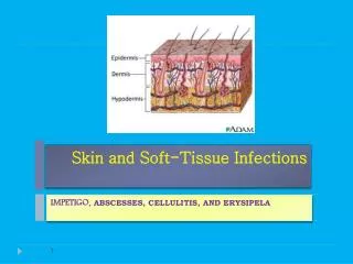
SKIN INFECTIONS. BACTERIA VIRUSES FUNGI. NORMAL SKIN FLORA. The resident flora of skin is a mixture of harmless staphylococcus epidermidis, corynebacterium spp.,propionibacterium spp.,brevibacteium spp., malassezia spp. In a minority, carry it in their nostrils, perineum or armpits.

E N D
BACTERIA • VIRUSES • FUNGI
NORMAL SKIN FLORA • The resident flora of skin is a mixture of harmless staphylococcus epidermidis, corynebacterium spp.,propionibacterium spp.,brevibacteium spp., malassezia spp. • In a minority, carry it in their nostrils, perineum or armpits.
Bacterial infections PYODERMAS : • Infections of skin and appendages commonly by S.aureus,Strep.pyogenes or both. • S.Aureus Nonfollicular- Bullous impetigo, impedigo contagiosa*, ecthyma* Follicular- superficial folliculitis, deep folliculitis, furuncle, carbuncle • Strep.pyogenes Localized- impetigo contagiosa*,ecthyma* Spreading- erysipelas, cellulitis *-Caused by one and/or both the organigms
Impetigo Types: impetigo contagiosa Bullous impetigo Causes • Staphylococci, streptococci, or by both together • Bullous type is usually caused by Staphylococcus aureus • The crusted ulcerated type is caused by β- haemolytic strains of streptococci. • Highly contagious
impetigo contagiosa • Epidemiology: • Prevalence: Frequent, often occuring in epidemics. • Age :preschool and young school children • Gender:adults,male>females • Clinical features: • Thin walled bulla on an erythematous base , ruputres rapidly to form an exudative plaque covered with honey –colored crusts.The lesion spreads peripherally without central healing and many lesions may coalesce to form polycyclic plaque.Removal of the crust reveals an erosion. • Usually multiple lesions, particularly around the face. • Regional lympadenopathy is frequent, • Constitutional symptoms may occur • Complications: Acute glomerulonephritis
Bullous impetigo • Epidemiology: Sporadic • Age: new borns and infants • Clinical features: • Bullae, containing turbid fluid, without an erythematous halo,rupture after few days to form thin, varnish like crusts.Lesion may heal in the center to form annular plaques. • Mucous membranes may be involved. • Site: face and other parts • Complications: • lymphadenopathy • staphylococcal scalded skin syndrome
Differential diagnosis • Herpes simplex • Eczema • Scalp lice • Bullous/contagiosum
Investigation • Diagnosis is usually made on clinical grounds • Gram stains or swabs are sent to the laboratory for culture, but treatment must not be held up until the results are available.
treatment • General measures: • local hygiene,gentle removal of the crust after softening with topical agent and compressing thems and application of a topical antibiotic such as neomycin, fusidic acid mupirocin or bacitracin is effective. • Systemic antibiotics (such as flucloxacillin,amoxy-clavulanic acid, erythromycin or cefalexin) are needed for severe cases • If a nephritogenic strain of streptococcus is suspected (penicillin V).
Furunculosis (boils) • A furuncle is an acute pustular infection of a hair follicle, usually with Staphylococcus aureus
Presentation and course • A tender red nodule which enlarges • Later may discharge pus • Its central ‘core’ before healing leave a scar • Site-face, axillae, buttocks and perineal region • Most patients have one or two boils • The sudden appearance of many furuncles suggests a virulent staphylococcus including strains of community- acquired MRSA.
chronic furunculosis • Susceptibility of follicles or colonization of nares or groins by pathogenic bacteria.
Predisposing factors • Nasal or perineal carriage • Diabetes,hiv • Underlying skin disease-scabies, atopic dermatitis
Complications • Septicemia
Differential diagnosis • Based on clinical diagnosis • Hidradenitis suppurativa should be considered if only the groin and axillae are involved.
Investigations in chronic furunculosis • General examination: look for underlying skin disease (e.g. scabies, pediculosis, eczema). • Test the urine for sugar. • Full blood count. • Culture swabs from lesions and carrier sites (nostrils, perineum) of the patient and immediate family. Test both to identify the organism and to evaluate sensitivity to various antibiotics. • Immunological evaluation only if the patient has recurrent or unusual internal infections too.
Treatment • Acute episodes- • moist hot fomentation,topical antibiotics • simple incision and drainage. • An appropriate systemic antibiotic is needed when • Many furuncles are erupting • When fever is present or • When the patient is immunosuppressed • Daily bath using an antiseptic soap. • Improve hygiene and nutritional state • Chronic: antibiotics topical and systemic
Staphylococcus Scalded skin syndrome(ssss) • Erythema and tenderness are followed by the loosening of large areas of overlying epidermis • In children, the condition is usually caused by a hematological spread of exotoxin produced by staphylococcal infection elsewhere (e.g. impetigo or conjunctivitis or otitis)which cause split in upper layers of epidermis. • No mucosal involvement • Good prognosis
In adults with widespread exfoliation, consider toxic epidermal necrolysis Staphylococcal scalded skin syndrome in a child. The overlying epidermis is loosening in the red areas
Investigation and Treatment • Gm stain and culture • Supportive and nursing care • Systemic antibiotics iv then later oral drugs(antistaph.)
Carbuncle • A group of adjacent hair follicles becomes deeply infected with Staphylococcus aureus • Swollen painful suppurating area of discharging pus from several points. Commonest site back and neck and thigh • The pain and systemic upset are greater than those of a boil. • Diabetes and Sys.steroid therapy must be excluded and Pus culture should be done • Treatment needs both topical and systemic antibiotics(flucloxacillin or other penicillinase resistant antiiotics). • Incision and drainage have been shown to speed up healing, although it is not always easy when there are multiple deep pus-filled pockets. • Consider the possibility of a fungal kerion in unresponsive carbuncles.
Toxic shock syndrome • Life threatening disease • A staphylococcal toxin is also responsible for this condition • Mostly in females during or imediately after menstuation and is associated with use of highly absorbent intravaginal tampons. • High fever , systemic upset (myalgia, headache, sore throat and vomiting),generalised erythematous blancing rash and hypotension • Multisystem involvement withen hrs • Erythema – and sometimes circulatory collapse • A week or two later- characteristic desquamation, most marked on the fingers and hands. • Immediate resuscitation ,systemic antibiotics (fluclox or vancomycin) and irrigation of the underlying infected site are needed.
Streptococcal infections • Erysipelas • Cellulitis • Necrotizing fasciitis
Erysipelas • It is superficial spreading puoderma while cellulitis is deeper and often the 2 coesist. • The causative streptococci usually gain their entry through a split in the skin (e.g. between the toes or under an ear lobe). • The first warning of an attack • Malaise • Shivering • Fever • After a few hours the affected area of skin becomes • red, and the eruption spreads with a well-defined advancing edge. • Blisters may develop on the red plaques. • Untreated, the condition can even be fatal. • It responds rapidly to systemic penicillin.
Cellulitis • This inflammation of the skin occurs at a deeper level than erysipelas. • The subcutaneous tissues are involved and the area is more raised and swollen. • The erythema is less marginated than in erysipelas. • Cellulitis often follows an injury,most common site is lower limbs • Favours areas of hypostatic oedema. • Streptococci, staphylococci or other organisms may be the cause. • Treatment is elevation, rest,NSAIDS – sometimes in hospital • Systemic antibiotics-Penicillin(erythromycin),if recurrent chemoprophylacis with long acting penicillin (benzathine penicillin)
Leprosy • Leprosy (Hansen's disease) is a chronic granulomatous disease affecting skin and nerve. • Causative organism: Mycobacterium leprae(AFB) • Infection: • nasal droplets followed by haematogenous spread to skin and nerve. • The clinical features depend on the immunological status of the host and not on the virulence of the organism : good cell mediated response -localized infection poor immunological response -generalized inf.of skin along with visceral involvement seen.
Pathogenesis • At the tuberculoid pole, • well-expressed CMI and delayed hypersensitivity control bacillary multiplication; organised epithelioid granulomas are seen in tissue biopsies. • Th1-type response to M. leprae, producing interleukin-2 (IL-2) and interferon-γ (IFN-γ), and positive lepromin (a soluble M. leprae preparation) skin tests. This strong cell-mediated response clears antigen, but with local tissue destruction. • In the lepromatous form, there is abundant bacillary multiplication in Schwann cells and the perineurium. • have a specific cell-mediated T-cell and macrophage anergy to M. leprae and poor lymphocyte responses to M. leprae antigens in vitro. They are negative on lepromin skin testing. They produce Th2-type cytokines. Nerve damage occurs across the spectrum in skin lesions and peripheral nerves. • Between these two poles is a continuum, varying from patients with moderate CMI (borderline tuberculoid) to patients with little cellular response (borderline lepromatous).
Classification • Lepromatous • Borderline lepromatous(BL) • Borderline • Borderline tuberculoid(BT) • Tuberculoid • CARDINAL FEATURES OF LEPROSY • Skin lesions, typically anaesthetic at tuberculoid end of spectrum • Thickened peripheral nerves • Acid-fast bacilli on skin smears or biopsy
Presentation • Lepromatous leprosy Skin • macules numerous, hypopigmented and erythematous or plaques, papules, or diffuse infiltration of the skin occur. • In advanced cases: nodular lesion especially on ears face and eyebrows are lost • Leonie facies-thickened brow and lobes of the ear Late features • Nerve enlargement and damage • Anaesthesia. In skin lesions the small dermal sensory and autonomic nerve fibres are damaged, causing local sensory loss (touch,heat,pain,numbness)and loss of sweating (anhydrosis)within that area. First in the distal aspects of the forearms and lower legs later ‘glove and stocking’distribution and eventually over the trunk and face. • Weakness and wasting of muscles of hands feet and face
Testes atropy, impotence, gynaecomastia • Mucosa of nose mouth pharynx and larynx:rhinitis hoarsenes, perforationof nasal septum and palate larygeal obstruction. • Bones of hands feet and face: cystic lesions of phalanges permitting fracture loss of upper incisor teeth and nasal spine leaking to nasal collapse • Eye involvement. Blindness due to leprosy is a devastating complication for a patient with anaesthetic hands and feet. Eyelid closure is impaired when the facial (7th) nerve is affected. Damage to the trigeminal (5th) nerve causes anaesthesia of the cornea and conjunctiva. The cornea is then susceptible to trauma and ulceration.
Tuberculoid leprosy: Depigmentation with a palpable erythematous rim at the upper edge. The ‘leonine’ facies of lepromatous leprosy
Presentation • Tuberculoid leprosy • one or few solitary lesions in skin and peripheral nerves Skin lesions • Macular or raised as plapues or as rings with flat center • The lesion is hypopigmented in dark skins coppery in pale skins • Well defined margin surface is dry and scaly • Lesion are of any size and occur any where • Loss of sensation is early and marked Peripheral neuropathy. • Thickened nerves, sensory loss and dysfucn of muscles(asthenia)supplied by that peripheral nerve • 'sites of predilection'. ulnar (above elbow)nerve-claw hand • median (wrist or above elbow), radial (humerus) -wrist drop, • radial cutaneous (wrist),common peroneal (knee), posterior tibial • facial nerve as it crosses the zygomatic arch, and great auricular in the posterior triangle of the neck.
Borderline or dimorphous(BLand BT)leprosy • Lesion are intermediate in character betn lepromatous and tubrculoid or as a mixture of them • Borderline leprosy is unstable • Borderline lepromatous leprosy (BL) is characterised by widespread small macules. They may experience both type 1 and type 2 reactions. Peripheral nerve involvement is widespread. • Borderline tuberculoid (BT) The skin lesions are similar to those in tuberculoid leprosy but are more numerous. Damage to peripheral nerves may be widespread and severe. These patients are prone to type 1 reactions with consequent nerve damage. • Pure neural leprosy This occurs principally in India and accounts for 10% of patients. There is asymmetrical involvement of peripheral nerve trunks and no visible skin lesions. On nerve biopsy all types of leprosy have been found.
Leprosy reactions • Leprosy reactions are events superimposed on the cardinal features .Type 1 (reversal) reactions These occur in 30% of borderline patients (BT, BB, BL) and are delayed hypersensitivity reactions caused by increased recognition of M. leprae antigens in skin and nerve sites. • Type 2 (erythema nodosum leprosum-ENL) reactions These are partly due to immune complex deposition and occur in BL and LL patients who produce antibodies and have a high antigen load.
REACTIONS IN LEPROSY • Indicated for any new impairment of nerve or eye function.1% hydrocortisone drops or ointment and 1% atropine drops.
Differential diagnosis • Skin The anaesthesia of tuberculoid and borderline tuberculoid lesions differentiates them from fungal pityriasis versicolor, vitiligo, post-inflammatory depigmentation, psoriasis and eczema. • The presence of acid-fast bacilli in smears differentiates lepromatous nodules from onchocerciasis, Kaposi's sarcoma and post-kala-azar dermal leishmaniasis. • Nerves Leprosy is the most common cause of peripheral nerve thickening. Uncommon conditions such as Charcot-Marie-Tooth disease and amyloid are differentiated from leprosy by the absence of skin lesions and acid-fast bacilli. Comparison should always be made with nerves on the other side. The causes of other polyneuropathies such as HIV infection, diabetes, alcoholism, vasculitides and heavy metal poisoning should all be considered where appropriate.
Investigations • The diagnosis is clinical, made by finding a cardinal sign of leprosy and supported by finding acid-fast bacilli in slit skin smears or typical histology in a skin or sensory nerve biopsy. • Skin lesions should be tested for anaesthesia. • The peripheral nerves should be palpated for thickening and tendernessand motor and sensory test to be done.CNS is unaffected. • Slit skin smears : The bacterial load is assessed by scraping dermal material on to a glass slide. The smears are then stained and acid-fast bacilli are scored on a logarithmic scale: the bacterial index (BI). Smears are useful for confirming the diagnosis and monitoring response to treatment. • Lepromin test. • This is of no use in the diagnosis of leprosy but, once the diagnosis has been made, it will help to decide which type of disease is present (positive in tuberculoid type).
Management • PRINCIPLES OF LEPROSY TREATMENT • Stop the infection with chemotherapy • Treat reactions • Educate the patient about leprosy • Prevent disability • Support the patient socially and psychologically • All leprosy patients should be given an appropriate multidrug combination. Patients can be classified into paucibacillary (skin smear-negative tuberculoid and BT) and multibacillary (skin smear-positive BT, all BB, BL and LL).Multibacillary patients with an initial BI>4 need longer treatment and should be treated to smear negativity(24mths)
MODIFIED WHO-RECOMMENDED MULTIDRUG THERAPY REGIMENS IN LEPROSY • WHO classification for field use when slit skin smears are not available • paucibacillary single-lesion leprosy (one skin lesion) • paucibacillary (2-5 skin lesions) • multibacillary (more than 5 skin lesions).In this field classification WHO recommends treatment of multibacillary patients for 12 months only.
Patient education: Reassurance that after 3 days of chemotherapy they are not infectious and can lead a normal social life. • Prevention of disability • Physiotherapy can prevent contractures, muscle atrophy and over-stretching of paralysed muscles. Anaesthetic feet need protective footwear. • Social, psychological and economic rehabilitation . • Prognosis The majority of patients, especially those who have no nerve damage at the time of diagnosis, do well on MDT, with resolution of skin lesions. Borderline patients are at risk of developing type 1 reactions which may result in devastating nerve damage. • Prevention and control case detection and providing MDT. BCG vaccination has been shown to give good but variable protection against leprosy.
Cutaneous Tuberculosis • Cutaneous tuberculosis- is highly variable in its clinical presentation, depending on the immunologic status of the patient and the route of inoculation of mycobacteria into the skin. • In most cases, the organism reaches the skin via lymphatic or hematogenous spread. • Exogenous inoculation cutaneous tuberculosis does occur.