Download
1 / 63
680 likes | 1.7k Vues
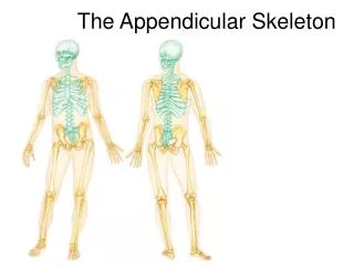
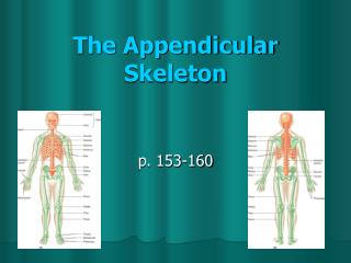
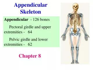
The Appendicular Skeleton. Chapter 8. The Appendicular Skeleton. Limb bones and their girdles are appended , or attached to the axial skeleton The pectoral girdle attaches the upper limbs to the trunk The pelvic girdle secures the lower limbs

E N D
The Appendicular Skeleton Chapter 8
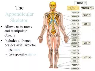
The Appendicular Skeleton • Limb bones and their girdles are appended, or attached to the axial skeleton • The pectoral girdle attaches the upper limbs to the trunk • The pelvic girdle secures the lower limbs • The upper and lower limbs differ in their functions but share the same structural plan
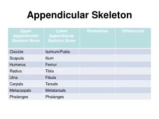
The Pectoral Girdle • Consists of the clavicle and the scapula - do not completely encircle the body • Medial end of each clavicle articulates with the manubrium and first rib • Laterally, the ends of the clavicles join the scapulae - scapulae do not join each other
Pectoral Girdle Functions • Provides attachment for many muscles that move the upper limb • The girdle is light allows upper limbs to be mobile • Only the clavicle articulates with the axial skeleton • Glenoid cavity - socket of the shoulder joint is shallow - good for flexibility but bad for stability
Clavicles (‘Little Keys’) • Aka collarbones are slender and S-shaped • Extend horizontally across the superior thorax • The flattened acromial end articulates with the scapula laterally • The cone-shaped sternal end attaches to the manubrium medially
Clavicle Functions • Provide attachment for muscles • Act as braces - holds the scapulae and arms out laterally from the thorax - a fractured clavicle will cause the entire shoulder region to collapse • Transmits compression forces from the upper limbs to the axial skeleton - allows you to push a heavy object
Scapulae • Are thin, triangular flat bones located on the dorsal surface of the rib cage - between rib 2 superiorly and rib 7 inferiorly • 3 borders: Superior – shortest and sharpest; Medial (vertebral) – parallels the vertebral column; Lateral (axillary) – abuts the axilla and ends superiorly in the glenoid cavity (shallow fossa) • 3 angles: Lateral – by the glenoid cavity; Superior – the superior and medial borders meet; Inferior – junction of the medial and lateral borders
Biceps muscle • Articulates with the humerus • Suprascapular nerve • Subscapularis muscle Fig 8.2
Muscles: • Infraspinatus • Supraspinatus Fig 8.2b
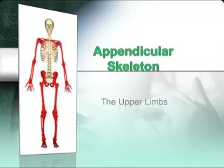
The Upper Limb • 30 bones – arm, forearm, and hand • Humerus – only bone of the arm - longest and strongest bone of the upper limb - articulates with the scapula at the shoulder - articulates with the radius and ulna at the elbow - provides sites for muscle attachment - provides articulation sites for other bones
Rotator cuff muscles • Guides a tendon of the biceps • Deltoid muscle • Radial nerve • Epicondyles muscle sites • ‘Pulley’ articulates with ulna • ‘Small head’ articulates with radius Fig 8.3
Antebrachium or Forearm • 2 parallel long bones, the radius and the ulna - articulate with the humerus proximally and bones of the wrist distally - articulate with each other proximally and distally at the radioulnar joints • Interosseous membrane interconnects the radius and ulna • In anatomical position the radius is lateral - when the palm faces posteriorly, the 2 bones form an X distal end of the radius crosses over the ulna
Details of Arm and Forearm a) Anterior view Fig 8.5a
Ulna (‘elbow’) • Main bone forming the elbow joint with the humerus - slightly longer than the radius, looks like a monkey wrench • 2 projections at the proximal end - olecranon process and coronoid process - separated by the trochlear notch (a deep concavity) • Hinge joint allows forearm to bend upon the arm (flex), then straighten again (extend) • Distally ulna shaft ends in a knoblike head that articulates with the radius - head is separated from the carpals by fibrocartilage disc that plays little or no role in hand movement
Ulna - Proximal Part • Extended – olecranon process locks into the olecranon fossa of the humerus Flexed – coronoid process fits into the coronoid fossa of the humerus Articulates with ? Fig 8.5b
Radius (‘rod’) • Thin at its proximal end and wider at distal end - proximal head shaped like the end of a thread spool - concave superior surface articulates with the capitulum of the humerus - medially radius head articulates with the ulna radialnotch to form the proximal radioulnar joint (__ joint?) - radial tuberosity, site of attachment for the biceps - styloid process, anchors a ligament - runs to the wrist • Primary forearm bone contributing to the wrist joint - when the radius rotates, the hand moves
Hand • Includes the following bones: Carpus – wrist Metacarpals – palm Phalanges – fingers • Carpus forms the true wrist – proximal region of the hand (just distal to the wrist joint) • Gliding movements occur between the carpals - make the wrist flexible
Carpus • Composed of 8 marble-sized bones • Proximal row, from lateral to medial: Scaphoid (‘boat-shaped’); Lunate (‘moonlike’) Triquetrum (‘triangular’); Pisiform (‘pea-shaped) - scaphoid and lunate bones articulate with the radius • Distal row from lateral to medial: Trapezium (‘little table’); Trapezoid (‘4-sided’) Capitate (‘head-shaped’); Hamate (‘hooked’) Sally Left The Party To Take Carmen Home
Bones of the Hand Fig 8.7
Metacarpus • 5 metacarpals radiate distally from the wrist to form the metacarpus or palm • Metacarpals form the palm: numbered I – V - beginning with the pollex - proximally bases articulate with the distal row of carpals - distally, bulbous heads articulate with the proximal phalanges
Phalanges • Numbered I – V, beginning with the pollex • Except for the thumb, each finger has 3 phalanges (miniature long bones): Proximal, Middle (missing in the thumb), and Distal
Pelvic Girdle • Attaches the lower limbs to the spine • Supports the visceral organs of the pelvis • Full weight of the upper body passes through the girdle - attaches to the axial skeleton by strong ligaments • Acetabulum socket - a deep cup that holds the head of the femur - lower limbs have less freedom of movement but are more stable than the arm
Pelvic Girdle • Consists of paired coxal (hip) bones or os coxae (os = bone; coxa = hip) • Hip bones unite anteriorly with each other and articulate posteriorly with the sacrum • Bony pelvis - deep, basinlike structure formed by the coxal bones, sacrum, and coccyx
Bony Pelvis Fig 8.8
Coxal Bones • Consist of 3 separate bones in childhood: - ilium, ischium, and pubis • Bones fuse but separate names identify regions of the coxal bones • Acetabulum – a deep hemispherical socket on the lateral pelvic surface
Large, flaring bone Forms the superior region of the coxal bone Site of attachment for many muscles Articulation with the sacrum forms the sacroiliac joint Bone Markings: - Ala (‘wing’) - Ilium crest - Ant sup/inf iliac spine - Pos sup/inf iliac spine - Greater sciatic notch - Pos, ant, inf gluteal lines - Iliac fossa - Auricular (‘ear’) surface – articulates with sacrum - Arcuate (‘bowed’) line Ilium
Ishium • Forms posteroinferior region of the coxal bone • Anteriorly – joins the pubis • Ischial tuberosities – strongest part of the hip bones • Superior ischium body bone markings: Ischial spine: attaches the scarospinous ligament Lesser sciatic notch: nerves and vessels for the perineum
Pubis or Pubic Bone • V- shaped (‘sexually mature’), with superior and inferior rami extending from a flat body - anterior border thickened to form a pubic crest - forms the anterior region of the coxal bone - lies horizontally in anatomical position • Pubic symphysis – pubic bones joined by fibrocartilage at the midline • Bone markings: Pubic tubercle – attachment point for the inguinal ligament Obturator foramen: between the pubis and ischium
True and False Pelves • Bony pelvis is divided into 2 parts: false and true - separated by the pelvic brim, a continuous oval ridge • False (greater) pelvis – superior to the pelvic brim - bounded by alae of the iliac bones • True (lesser) pelvis – inferior to pelvic brim - forms a bowl containing the pelvic organs
Pelvic Structures and Childbearing • Major differences between male and female - female pelvis adapted for childbearing - pelvis is lighter, wider, and shallower - provides more room in the true pelvis
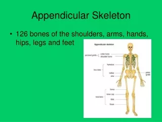
The Lower Limb • Carries the entire weight of the erect body • Bones of lower limb are thicker and stronger than those of upper limb • Divided into 3 segments: the thigh, leg, and foot
Thigh • Region of the lower limb between the hip and the knee • Femur – the single bone of the thigh - longest and strongest bone of the body - ball-shaped head articulates with the acetabulum • Bone markings: - Fovea capitis (‘pit of the head’): ligament of the head of the femur runs to the acetabulum - Greater and lesser trochanter: sites of muscle attachment - Gluteal tuberosity, linea aspera – muscle attachment sites - Lateral and medial epicondyles – muscles and ligaments
Patella • Triangular sesamoid bone • Imbedded in the tendon that secures the quadriceps muscles • Protects the knee anteriorly • Improves leverage of the thigh muscles across the knee
Leg • Refers to the region of the lower limb between the knee and the ankle - composed of the tibia and fibula • Tibia – more massive medial bone of the leg - receives weight of the body from the femur • Fibula – stick-like lateral bone of the leg • Interosseous membrane - connects the tibia and fibula
Tibia and Fibula • Tibia articulates with the femur at superior end - forms the knee joint • Tibia articulates with the talus at the inferior end - forms the ankle joint • Fibula does not contribute to the knee joint - stabilizes the ankle joint
The Foot • Composed of the tarsus, metatarsus, & phalanges • Functions: - supports body weight - acts as a lever to propel body forward when walking - segmentation makes foot pliable and adapted to uneven ground